Understanding The Intrinsic Rate Of The Sinoatrial Node: A Guide To Heartbeat Regulation

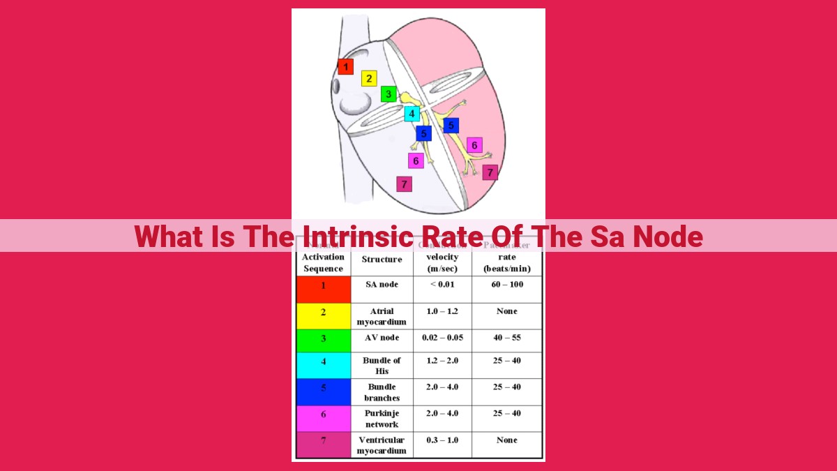
The intrinsic rate of the sinoatrial (SA) node is the inherent heartbeat speed generated by the SA node, the natural pacemaker of the heart. This rate is crucial for maintaining a regular cardiac rhythm and supplying the body with adequate blood flow. The SA node’s electrical impulses travel through specialized conduction pathways, including the atrioventricular (AV) node, bundle of His, and Purkinje fibers, ensuring coordinated heart contractions. The rate is influenced by ion channels that control the flow of ions across cell membranes, resulting in the cardiac action potential with phases of depolarization and repolarization.
- Explain the importance of the intrinsic rate of the SA node in maintaining the heart’s regular rhythm.
The Heart’s Rhythm: The Intrinsic Symphony of the SA Node
The human heart, a marvel of biological engineering, beats tirelessly, maintaining the flow of life-sustaining blood throughout our bodies. This rhythmic symphony is orchestrated by a tiny group of cells within the heart, known as the sinoatrial (SA) node.
The SA node, nestled in the right atrium, is the heart’s natural pacemaker. Its intrinsic rate determines the tempo of our heartbeat, ensuring a steady and consistent rhythm. This intrinsic rate is crucial for maintaining the heart’s normal function and preventing potentially life-threatening arrhythmias. If the SA node loses its ability to set the heartbeat, other heart cells can develop abnormal rhythms, leading to serious health complications.
Nodal Cells and the Electrical Conduction System of the Heart
Your heart is a remarkable organ, beating tirelessly throughout your life to pump blood and sustain your very existence. This rhythmic beating is orchestrated by a complex electrical system, and at its core lies the sinoatrial (SA) node, the heart’s natural pacemaker.
The SA node is a tiny cluster of cells located in the right atrium. These specialized cells possess the unique ability known as automaticity, generating electrical impulses spontaneously and at a regular rate. These impulses then spread through the heart’s conduction system, ensuring a coordinated contraction of the atria and ventricles.
The electrical impulses from the SA node first travel to the atrioventricular (AV) node, another collection of specialized cells located at the junction between the atria and ventricles. The AV node serves as a gatekeeper, delaying the impulses slightly to allow the atria to complete their filling before allowing them to pass through to the ventricles.
From the AV node, the impulses race down the bundle of His, a bundle of fibers that connects the AV node to the septum, the wall separating the left and right ventricles. The bundle of His then divides into left and right branches, delivering the impulses to the Purkinje fibers.
These Purkinje fibers are specialized muscle fibers that extend throughout the ventricles. They distribute the electrical impulses rapidly and evenly, ensuring that all parts of the ventricles contract simultaneously. This coordinated contraction is what generates the pumping action of the heart, propelling blood throughout your body.
The electrical conduction system, with its nodal cells and specialized fibers, is a masterpiece of nature’s engineering. It ensures the heart’s rhythmic and efficient beating, a vital process that sustains life and allows us to experience the wonders of the world.
The Rhythm of Life: Ion Channels and Membrane Potentials in the Heart
In the heart of the matter, the steady lub-dub sound is not just a simple beat; it’s a symphony of electrical impulses, meticulously conducted by specialized cells and ion channels. These channels, like gatekeepers, control the flow of charged particles, shaping the intricate electrical events that keep our hearts beating in rhythm.
Calcium and Potassium: The Conductors of Excitement
Among the symphony’s conductors are two key players: calcium (Ca2+) and potassium (K+) ions. Calcium, like a surge of energy, rushes into the cells, initiating the electrical impulse. Potassium, on the other hand, acts as a calming force, flowing out of the cells to restore balance.
The Orchestral Action: Membrane Potentials and the Cardiac Action Potential
As calcium and potassium ions conduct their dance, they create electrical potential differences across the cell membrane. This is known as the membrane potential. The heart’s electrical impulse, the cardiac action potential, is a series of these potential changes.
The action potential starts with a rapid depolarization, where calcium floods in, creating a positive charge inside. Next, repolarization sets in as potassium rushes out, restoring a negative charge. A final hyperpolarization overshoots the resting membrane potential, ensuring a complete reset before the next impulse.
These meticulously orchestrated ion movements, guided by the specialized ion channels, create the electrical rhythm that pumps life through our bodies.
Electrical Events:
- Describe the phases of the cardiac action potential, including hyperpolarization, depolarization, and repolarization.
Electrical Events: The Heart’s Rhythmic Dance
Within the heart’s intricate chambers, an intricate dance of electrical impulses unfolds, orchestrating the organ’s rhythmic beat. This dance, known as the cardiac action potential, is the foundation of the heart’s vital role in sustaining life.
Hyperpolarization: The Prelude to Excitation
The heart’s dance begins with hyperpolarization, a tranquil phase where resting potential reigns supreme. An influx of potassium ions swings the membrane potential deep into negative territory, creating a “resting” state of electrical silence.
Depolarization: The Spark of Excitation
But this tranquility is short-lived. A sudden surge of calcium ions rushes into the myocardial cells, triggering a dramatic shift in membrane potential. This rapid depolarization marks the onset of the action potential, the spark that ignites the heart’s electrical impulses.
Repolarization: The Return to Equilibrium
At the peak of depolarization, a swift reversal occurs. Potassium channels open wide, allowing a flood of potassium ions to flow out, repolarizing the membrane. The heart momentarily returns to its resting state.
Refractory Period: A Safeguard against Chaos
Following depolarization, the heart enters a brief but crucial refractory period, a time when it is temporarily more resistant to further excitation. This refractory period prevents the heart from repolarizing too quickly, safeguarding against potentially life-threatening arrhythmias.
Through this intricate symphony of electrical events, the heart’s rhythm is maintained, ensuring the steady flow of blood throughout the body. This rhythmic dance is a testament to the heart’s unwavering commitment to sustain life, a testament to its remarkable artistry.
Automaticity and Heart Rate: The Rhythm of Life
Nestled within the heart’s chambers lies a remarkable collection of cells known as the sinoatrial (SA) node. These cells possess a unique property called automaticity, the ability to generate electrical impulses spontaneously and rhythmically. This intrinsic rate of the SA node serves as the heart’s natural pacemaker, orchestrating the heartbeat that sustains our very existence.
Unveiling the Automated Cells
The SA node comprises specialized pacemaker cells that exhibit a constant depolarization known as the “pacemaker potential.” Unlike regular heart cells, which require an external stimulus to initiate depolarization, pacemaker cells generate their own electrical impulses due to a gradual influx of calcium and sodium ions through specific channels in their cell membranes.
Orchestrating the Heartbeat
The heart’s electrical system is a finely orchestrated symphony of electrical signals. Once generated in the SA node, the electrical impulse travels through the heart’s conducting system, which includes the atrioventricular (AV) node, bundle of His, and Purkinje fibers. Each component plays a crucial role in coordinating the timing and sequence of electrical events, ensuring the heart’s chambers contract in a synchronized manner.
Influencing Heart Rate
While the SA node establishes the intrinsic heart rate, numerous factors can modify it to meet the body’s ever-changing demands. The autonomic nervous system, comprised of the parasympathetic and sympathetic branches, exerts a profound influence on heart rate. Parasympathetic stimulation via the vagus nerve slows the heart, whereas sympathetic activation via catecholamines like adrenaline accelerates it.
Hormones such as thyroid hormones and epinephrine also impact heart rate. Thyroid hormones increase the rate of calcium influx into pacemaker cells, thereby increasing heart rate. Epinephrine, released during the body’s fight-or-flight response, directly binds to beta-adrenergic receptors on heart cells, leading to tachycardia (increased heart rate).
In essence, automaticity is the heart’s built-in rhythm generator, while external influences fine-tune the tempo to meet the body’s needs. By understanding the complexities of automaticity and its regulation, we can better appreciate the remarkable intricacies of our cardiovascular system, the unceasing engine driving life’s journey.