Comprehensive Tissue Processing And Analysis For Accurate Disease Diagnosis

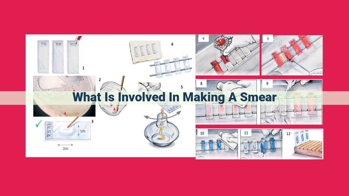
Making a smear involves preparing slides by processing, embedding, and sectioning tissues; collecting samples via biopsy, excision, or aspiration; staining tissues using histochemistry, immunohistochemistry, or FISH to highlight cellular structures; examining slides using various microscopy techniques; and interpreting results through histopathology, cytopathology, or molecular pathology to identify cellular abnormalities and diagnose diseases.
Slide Preparation: Laying the Foundation for Cellular Exploration
The Journey Begins: Tissue Processing and Embedding
Every histological journey begins with carefully processing the tissue to preserve its delicate structures. This meticulous procedure involves removing excess fluids, infiltrating the tissue with a stabilizing embedding medium, and solidifying it into a block called a paraffin block.
Slicing the Histological Landscape: Sectioning
Once embedded, the paraffin block is sliced into thin sections using a microtome. These sections provide a representative cross-section of the tissue, allowing us to visualize its cellular components under a microscope.
Sample Collection: Unveiling the Cellular Landscape
In the realm of diagnostic pathology, obtaining tissue samples is a crucial step that ensures the accurate identification and characterization of cellular abnormalities. This process, known as sample collection, involves a variety of techniques tailored to the specific needs of each patient and the suspected disease.
One common method for collecting tissue samples is biopsy. This procedure involves removing a small piece of tissue from the suspected area using a specialized needle or scalpel. Biopsies can be performed in a variety of settings, including a doctor’s office, clinic, or hospital.
Excision is another technique used to collect tissue samples. In this procedure, the entire suspected lesion or growth is surgically removed and sent to the laboratory for examination. Excision is often used when a biopsy is not feasible or when a larger sample is needed for more thorough analysis.
In some cases, tissue samples can be collected using aspiration. This technique involves inserting a thin needle into the suspected area and using suction to remove a sample of cells. Aspiration is commonly used for collecting samples from lymph nodes, body fluids, and other areas where a biopsy or excision may not be practical.
Regardless of the method used, the goal of tissue sample collection is to obtain a representative specimen that accurately reflects the cellular characteristics of the suspected lesion. This ensures that pathologists have the necessary material to identify cellular abnormalities, diagnose diseases, and guide patient treatment.
Staining Techniques: Illuminating Cellular Structures
When examining tissue slides under a microscope, staining techniques play a crucial role in revealing intricate cellular details and facilitating accurate diagnoses. These techniques employ a variety of dyes and reagents to highlight specific tissue components and visualize their interactions, shedding light on the underlying cellular landscape.
Histochemistry: Unmasking Tissue Chemistry
Histochemistry utilizes chemical reactions to reveal the presence and distribution of specific molecules within cells and tissues. These reactions produce distinctive colors that correspond to the target molecules, allowing pathologists to identify and characterize cellular structures.
For instance, hematoxylin and eosin (H&E) staining is a common histochemical technique that stains cell nuclei blue and cytoplasmic components pink, providing a basic overview of tissue morphology. Other histochemical stains can target specific molecules, such as collagen, carbohydrates, or lipids, offering insights into tissue composition and function.
Immunohistochemistry: Pinpointing Protein Expression
Immunohistochemistry (IHC) employs antibodies to specifically bind to target proteins within cells. These antibodies are labeled with colored dyes, allowing researchers and pathologists to visualize the expression and localization of specific proteins.
IHC has revolutionized the field of pathology by enabling the identification of cellular markers associated with various diseases. For example, IHC can detect the presence of hormone receptors in breast cancer, aiding in targeted therapies and prognostication.
FISH: Illuminating Genetic Abnormalities
Fluorescence in situ hybridization (FISH) is a powerful technique that uses fluorescently labeled DNA probes to detect specific gene sequences within cells. These probes bind to complementary DNA sequences on chromosomes, illuminating genetic abnormalities such as translocations, deletions, or amplifications.
FISH is particularly valuable in diagnosing genetic disorders, such as Down syndrome or certain types of cancer. By identifying chromosomal aberrations, FISH provides crucial information for genetic counseling and targeted treatment strategies.
Staining techniques are indispensable tools in the arsenal of microscopy, enabling researchers and pathologists to unravel the hidden complexities of cells and tissues. From histochemistry to immunohistochemistry to FISH, these techniques provide a gateway to understanding cellular function, diagnosing diseases, and guiding personalized treatments. By illuminating cellular structures and interactions, staining techniques empower us to delve into the microscopic world and uncover the secrets that shape life and health.
Microscopic Examination: Unveiling Cellular Details
Microscopic examination lies at the heart of histopathology, illuminating the intricate world within our cells and tissues. Through various microscopy techniques, pathologists embark on a journey of discovery, seeking clues to unravel the mysteries of health and disease.
One of the oldest and most widely used techniques is light microscopy. This workhorse of histopathology allows pathologists to examine thin tissue sections under a microscope using visible light. By employing specialized stains or dyes, different structures within cells and tissues become visible, revealing their shapes, sizes, and relationships.
For even finer details, pathologists turn to electron microscopy. This powerful technique utilizes a beam of electrons to generate highly magnified images, allowing them to delve into the ultrastructure of cells and tissues. Electron microscopy unveils previously hidden organelles, membranes, and molecular components, providing invaluable insights into cellular processes and abnormalities.
Another indispensable tool in the microscopist’s arsenal is fluorescence microscopy. This technique harnesses the power of fluorescent dyes or antibodies that emit light of specific wavelengths when illuminated. By employing fluorescence microscopy, pathologists can visualize specific proteins, genetic material, or other molecules of interest, offering precise information about cellular localization and function.
Each microscopy technique has its unique strengths and applications. Light microscopy provides a broad overview of tissue architecture and cellular morphology. Electron microscopy offers ultrastructural details for in-depth analysis of cellular components and abnormalities. Fluorescence microscopy enables targeted visualization of specific molecules, allowing for highly specific and sensitive detection. The combination of these techniques empowers pathologists to obtain a comprehensive understanding of cellular events at multiple scales.
Unraveling the Diagnosis: Interpreting Slide Results
In the world of medical diagnostics, slides hold the key to unlocking the secrets hidden within the human body. Through a series of meticulous processes, tissue samples are transformed into slides that undergo microscopic scrutiny, revealing intricate cellular structures and subtle abnormalities. The interpretation of these slides plays a pivotal role in diagnosing diseases, enabling clinicians to make informed decisions about patient care.
Histopathology, cytopathology, and molecular pathology are essential disciplines that delve into the intricacies of slide interpretation. Histopathology examines tissue sections, providing insights into the cellular architecture and tissue organization. Cytopathology, on the other hand, focuses on individual cells, identifying abnormalities that may indicate disease. Molecular pathology utilizes advanced techniques to analyze the genetic makeup of cells, uncovering mutations and other genetic alterations linked to specific disorders.
The process of slide interpretation involves meticulous examination of the stained tissue or cell samples. Pathologists, highly trained medical professionals, pore over the slides, searching for telltale signs of cellular pathology. They assess cell morphology, noting changes in size, shape, and nuclear features. They also evaluate cell arrangement, looking for abnormal patterns that may indicate tissue invasion or malignancy.
Beyond routine microscopic examination, special stains and advanced technologies enhance the diagnostic capabilities of slide interpretation. Immunohistochemistry highlights specific proteins within cells, aiding in the identification of different cell types and the detection of disease-associated markers. Fluorescence in situ hybridization (FISH) visualizes specific DNA sequences, allowing for the detection of chromosomal abnormalities and genetic disorders.
The interpretation of slide results is not merely an exercise in pattern recognition; it requires a deep understanding of cellular pathology and a keen eye for detail. Pathologists must integrate their microscopic observations with clinical information, such as the patient’s history and physical examination findings, to formulate an accurate diagnosis. This interplay between microscopic analysis and clinical context is essential for guiding patient management and ensuring optimal care.
Through the meticulous interpretation of slide results, medical professionals gain invaluable insights into the cellular landscape of disease. Histopathology, cytopathology, and molecular pathology empower clinicians to make well-informed decisions, leading to timely interventions and improved patient outcomes.