Radial Fibers In Cell Division: Key Components Of The Mitotic Spindle

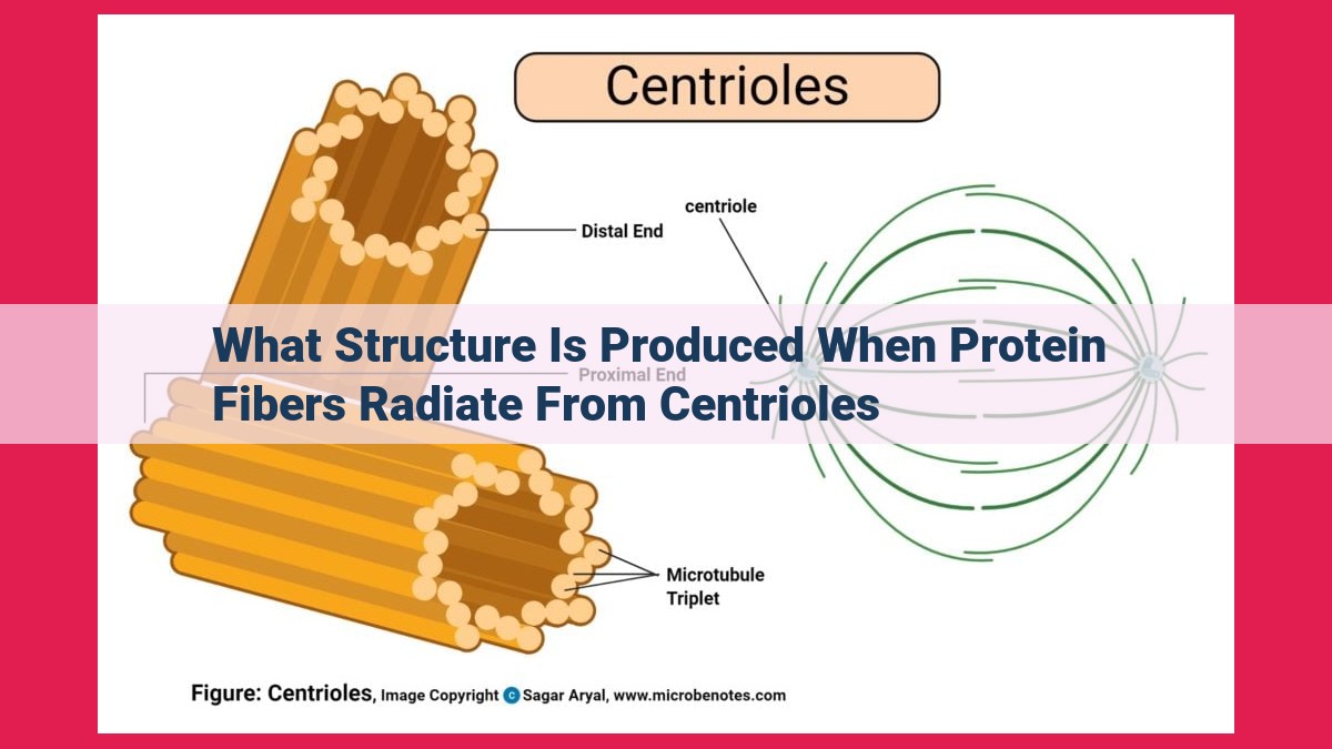
During cell division, protein fibers called radial fibers extend from centrioles, the organizers of spindle fibers. These fibers create the mitotic spindle, a structure that separates chromosomes during cell division. Centrioles are located at the spindle poles and serve as microtubule organizing centers, directing the growth of spindle fibers. Radial fibers interact with spindle fibers to define the spindle’s shape and contribute to its stability, ensuring accurate chromosome segregation and genetic integrity.
The Mitotic Spindle: The Orchestrator of Cell Division
In the intricate world of cellular biology, the mitotic spindle takes center stage as the maestro of cell division. This intricate apparatus governs the precise movement of chromosomes, ensuring their equitable distribution during cell division. At the heart of this process lie centrioles, the enigmatic organelles that orchestrate the spindle’s construction.
Centrioles, tiny cylindrical structures found near the nucleus, are the master architects of the mitotic spindle. They act as microtubule organizing centers, generating microtubules, the building blocks of the spindle. As they duplicate, centrioles form the poles of the spindle, from which radial fibers extend outward like spokes of a wheel. These fibers help determine the spindle’s shape and stability, ensuring the chromosomes’ proper alignment.
Spindle fibers, the core components of the spindle, connect the centrioles and provide the force necessary for chromosome movement. They attach to specialized structures called kinetochores on the chromosomes, guiding their precise separation and distribution.
Centrioles: The Unsung Heroes of Cell Division
Imagine a grand orchestra, its instruments harmoniously working together to produce a beautiful symphony. In the realm of cell division, that grand orchestra is the mitotic spindle apparatus, responsible for the meticulously orchestrated dance of chromosomes. And within this intricate machinery, the centrioles play a pivotal role, akin to the skilled conductors who guide the ensemble.
Structure and Location
Centrioles are cylindrical structures, found in pairs near the nucleus of animal cells. Their presence signals the onset of cell division, as they become the focal point for the organization of microtubules, the building blocks of the mitotic spindle.
Microtubule Organizing Centers
Centrioles act as microtubule organizing centers (MTOCs). They generate and extend microtubules in a specific orientation, like a skilled threader guiding invisible strings. These microtubules form the foundation of the spindle fibers and radial fibers, which are crucial for chromosome segregation.
Radial Fibers
From each centriole, radial fibers radiate outward like spokes of a wheel. These fibers interact with spindle fibers to create the characteristic bipolar shape of the mitotic spindle. Together, they form a stable scaffold, providing the necessary tension for chromosome alignment and separation.
Microtubules: The Architectural Pillars of Cell Division
Microtubules, the workhorses of the cellular world, play an indispensable role in the intricate dance of cell division. These cylindrical structures, composed of tubulin proteins, are the building blocks of the spindle apparatus, a sophisticated machinery that ensures the equitable distribution of chromosomes to daughter cells.
Each microtubule is a hollow tube, resembling a tiny straw. It assembles from the polymerization of tubulin dimers, which align themselves in a head-to-tail fashion. This polarized structure gives microtubules a defined polarity, with plus and minus ends.
The plus ends of microtubules are the active sites, where subunits are added to extend the structure. In the context of cell division, these plus ends are oriented towards opposite poles of the cell, forming the spindle fibers that stretch between the centrioles.
Microtubules, working in concert, fulfill multiple functions in the mitotic spindle. They form the structural framework, ensuring the spindle’s shape and stability. Additionally, microtubules generate the force necessary for chromosome segregation. The attachment of kinetochores, protein complexes located on chromosomes, to the spindle fibers allows the orchestrated movement of chromosomes towards their designated poles.
Radial Fibers: Guardians of Mitotic Stability
As the mitotic spindle begins to take shape, a network of radial fibers extends from the centrioles like a spider’s web. These delicate structures play a crucial role in maintaining the spindle’s form and guiding the movement of chromosomes.
Originating from the centrioles, radial fibers radiate outward, interacting closely with the spindle fibers. This intricate dance not only maintains the spindle’s bipolar shape but also ensures its stability.
Radial fibers are essential for preventing the spindle from collapsing and for creating a stable framework that facilitates chromosome segregation. Without their guiding presence, cell division could go awry, potentially leading to genetic instability.
Additional Notes for SEO Optimization:
- Use relevant keywords: “radial fibers,” “mitotic spindle,” “chromosome segregation”
- Include subheadings for each section
- Optimize image alt tags: “Radial fibers extending from centrioles”
- Ensure readability with short paragraphs and clear language
Spindle Fibers:
Spindle Fibers: The Guiding Lights of Chromosome Segregation
The mitotic spindle apparatus is a complex machine that orchestrates the precise segregation of chromosomes during cell division. At the heart of this apparatus are delicate filaments known as spindle fibers. These fibers serve as a guiding force, ensuring that each newly divided cell receives the correct complement of genetic material.
Formation and Structure of Spindle Fibers
Spindle fibers originate from two centrosomes, which reside at opposite poles of the cell. These centrosomes contain centrioles, cylindrical structures that act as microtubule organizing centers (MTOCs). Centrioles produce numerous microtubules, which radiate outward from the poles to form the spindle fibers.
Interplay with Kinetochores and Chromosomes
Each spindle fiber is composed of two polar microtubules that extend towards the opposite poles. At the center of the spindle, these fibers meet and interact with a specific region on each chromosome called the kinetochore. The attachment of spindle fibers to kinetochores allows the chromosomes to align themselves at the equator of the spindle, ready for segregation.
Force Generation for Chromosomal Movement
Once the chromosomes are aligned, the spindle fibers begin to shorten through a process known as kinetochore fiber attachment. This shortening generates a pulling force that drives the chromosomes apart, towards opposite poles of the spindle apparatus. The precise coordination of spindle fiber shortening ensures that each newly divided cell inherits the correct number of chromosomes.
The Importance of Spindle Fibers for Cell Division
The proper formation and function of spindle fibers are crucial for the accurate segregation of chromosomes. Without these guiding filaments, the chromosomes would fail to divide evenly, leading to genomic instability and potentially fatal consequences for the cell. Therefore, the mitotic spindle apparatus, including its spindle fibers, plays an indispensable role in ensuring the faithful transmission of genetic material and the maintenance of genetic stability across generations.
Unraveling the Secrets of the Mitotic Spindle: A Journey into the Heart of Cell Division
Imagine a microscopic theater within your cells, where chromosomes perform a meticulously choreographed dance. The stage for this performance is the mitotic spindle, an extraordinary apparatus that orchestrates the precise division and distribution of genetic material.
Centrioles: The Conductors of Microtubule Orchestra
At the heart of the spindle lie centrioles, enigmatic structures that initiate the assembly of microtubules, the sinews that hold the chromosomes in place. Centrioles, like miniature windmills, spin out thin, rigid radial fibers that radiate outward from the poles of the spindle.
Microtubules: The Force Behind Chromosome Segregation
Microtubules are the workhorses of the spindle, forming its supporting framework. They align themselves along the equator of the cell, creating spindle fibers that connect to the kinetochores, specialized protein complexes on the chromosomes. These spindle fibers act as molecular tug-of-war teams, pulling chromosomes to opposite poles of the cell.
Radial Fibers: Shaping and Stabilizing
Together, the radial fibers and spindle fibers shape the mitotic spindle into a bipolar spindle, creating a stable platform for chromosome movement. The radial fibers, in their fan-like arrangement, contribute to the spindle’s rigidity, ensuring that the chromosomes are segregated accurately.
Aster: A Halo of Stability
Surrounding the spindle poles are asters, radiating microtubules that contribute to spindle stability and positioning. Like celestial beacons, the asters guide the spindle fibers towards their targets, ensuring that chromosomes are moved to the correct locations.
The Dance of Division
As the cell prepares to divide, centrioles begin to separate and migrate to opposite poles. The radial and spindle fibers assemble, forming the framework of the mitotic spindle. Kinetochores attach to the spindle fibers, connecting chromosomes to the spindle apparatus.
The spindle fibers then shorten, pulling the chromosomes to the poles of the cell. This process, known as anaphase, is the climax of cell division. Finally, the cell membrane pinches in, creating two daughter cells, each with its own set of chromosomes.
The Guardians of Genetic Stability
The mitotic spindle is a crucial player in cell division, ensuring the accurate and equitable distribution of genetic material. Defects in spindle assembly or function can lead to aneuploidy, a condition where cells have an abnormal number of chromosomes. This can have devastating consequences, including developmental abnormalities and cancer.
By unraveling the secrets of the mitotic spindle, scientists gain invaluable insights into the fundamental mechanisms that govern cell division. This knowledge not only deepens our understanding of cellular biology but also paves the way for novel therapies targeting spindle function in disease.
The Incredible Microtubule Orchestra: Unveiling the Secrets of Cell Division
Cell division is a crucial process that ensures the growth, repair, and reproduction of all living organisms. At the heart of this intricate dance lies the mitotic spindle apparatus, a highly organized network of microtubules that ensures the precise segregation of chromosomes.
Centrioles: The Orchestra’s Conductors
Nestled within the cell’s cytoplasm, centrioles are cylindrical structures that act as the organizers of the microtubule orchestra. They orchestrate the formation of radial fibers, which radiate outward from the centrioles, and spindle fibers, which connect the centrioles and guide chromosome movement.
Microtubules: The Musical Instruments
Composed of tubulin proteins, microtubules are rigid, hollow tubes that give the mitotic spindle its shape and function. They assemble and disassemble dynamically, providing the force necessary for accurate chromosome segregation.
Radial Fibers: The Supporting Scaffolding
Radial fibers extend outward from the centrioles and interact with spindle fibers, forming a framework that supports the mitotic spindle. They contribute to the spindle’s shape and stability, ensuring its proper orientation for chromosome segregation.
Spindle Fibers: The Choreographers
Spindle fibers connect the centrioles and extend towards the chromosomes. These fibers serve as the tracks along which chromosomes “dance” during segregation. They attach to specific proteins on the chromosomes, known as kinetochores, ensuring their proper alignment.
Mitotic Spindle: The Grand Stage
The mitotic spindle, composed of microtubules, radial fibers, and centrioles, forms a complex yet elegant structure that orchestrates the precise separation of chromosomes. Its function is critical for ensuring genetic accuracy and cell survival.
Aster: The Radiant Halo
At the poles of the mitotic spindle lies the aster, a radiating structure composed of microtubules. The aster contributes to the spindle’s shape and stability, providing a foundation for the microtubule orchestra’s performance.
The mitotic spindle apparatus is an intricate orchestra that plays a vital role in cell division. Centrioles, microtubules, radial fibers, spindle fibers, and the aster work harmoniously to ensure the precise segregation of chromosomes. This process is fundamental to the growth, development, and genetic stability of all living organisms.