Peroneus Longus: The Key Muscle For Foot Eversion And Ankle Stability

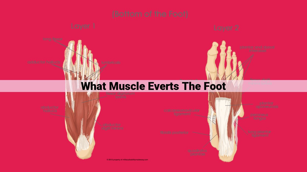
The peroneus longus muscle, originating at the lateral aspect of the fibula, is the primary everter of the foot. Its robust tendons course behind the lateral malleolus and insert on the lateral aspect of the base of the first metatarsal and medial cuneiform. Innervated by the superficial peroneal nerve, this muscle everts the foot, lifting its medial border and stabilizing the ankle joint. It’s assisted by the peroneus brevis, which inserts on the base of the fifth metatarsal, and the peroneus tertius, which originates from the tibia and inserts on the base of the fifth metatarsal. These muscles work synergistically to ensure proper foot eversion, maintaining balance and preventing excessive inward rolling of the foot.
Understanding the Muscles Involved in Foot Eversion
Foot eversion, the outward turning of the foot, plays a pivotal role in maintaining balance and stability. This movement is essential for activities that involve weight-bearing and coordination, such as walking, running, and jumping.
The muscles responsible for foot eversion are located in the lateral compartment of the lower leg. They work together to abduct (pull away from the midline) and evert (rotate outward) the foot. The primary evertor muscles are the peroneus longus, peroneus brevis, and peroneus tertius.
These muscles originate from the bones of the fibula and tibia and insert into the bones of the foot. They are innervated by the common fibular nerve. Together, they form a muscular “sling” that wraps around the outside of the ankle, providing stability and controlling the movement of the foot.
Meet the Peroneus Longus: A Key Everter
Origin:
The peroneus longus muscle arises from the lateral side of the fibula, a bone running alongside the tibia in your lower leg. Its origin spans from just below the knee down to the ankle.
Insertion:
From its origin, the peroneus longus travels laterally before crossing behind the lateral malleolus (bony prominence on the outer ankle) and inserting into the base of the first metatarsal (bone of the big toe).
Innervation:
The peroneal nerve, a branch of the common fibular nerve, supplies the peroneus longus muscle with motor innervation, allowing it to contract and move.
Function:
The primary role of the peroneus longus is foot eversion—rotating the sole of the foot outward. It works in conjunction with the peroneus brevis and peroneus tertius muscles to control the lateral (outer) movements of the foot. Additionally, it assists in dorsiflexion (lifting the foot upward) and pronation (flattening) of the foot during walking and running.
During eversion, the peroneus longus contracts, pulling the base of the first metatarsal laterally. This rotates the front of the foot outward, counteracting the natural tendency of the foot to roll inward (pronate) during weight-bearing. This everting action is essential for maintaining balance and stability while standing, walking, and running.
The Peroneus Brevis: A Loyal Assistant in Foot Eversion
Meet the Peroneus Brevis
In the intricate tapestry of foot muscles, the peroneus brevis plays a vital supporting role in the crucial movement of foot eversion. It’s an unsung hero that works alongside the stronger peroneus longus, providing assistance and ensuring smooth, coordinated eversion.
Origin and Insertion: A Tale of Two Bones
The peroneus brevis originates from the middle third of the lateral fibula, the long, slender bone on the outer side of the ankle. Its journey ends at the base of the fifth metatarsal, the long bone that forms the outermost toe. This arrangement allows it to effectively evert the foot, pulling it outward and away from the midline.
Innervation: A Nerve’s Guidance
The peroneal nerve, a branch of the sciatic nerve, commands the peroneus brevis, sending electrical impulses that trigger muscle contractions. This nerve connection ensures precise control and coordination with other leg muscles.
Function: The Art of Eversion
The peroneus brevis, along with its peroneus longus counterpart, plays a dominant role in everting the foot. Eversion is the outward and lateral movement of the foot, crucial for maintaining balance and stability while standing and walking. It allows us to push off and propel ourselves forward, as well as to navigate uneven surfaces with confidence.
Interplay with the Peroneus Longus: A Dynamic Duo
Together, the peroneus brevis and peroneus longus form a formidable alliance, collaborating seamlessly to evert the foot. The peroneus brevis assists the peroneus longus, providing additional force to overcome resistance and ensure efficient eversion. Their coordinated action prevents the foot from rolling inward (pronation), which can lead to potential injuries.
Peroneus Tertius: The Unsung Hero of Foot Eversion
In the intricate symphony of muscles orchestrating foot movement, there exists an often-overlooked player: the peroneus tertius. This unassuming muscle, nestled between its larger counterparts, plays a vital role in maintaining stability and balance.
Origin, Insertion, and Innervation:
The peroneus tertius originates from the lower third of the fibula, a long bone that runs alongside the tibia in the leg. It inserts onto the dorsal surface of the fifth metatarsal bone, the bone forming the outside edge of the foot. The muscle is innervated by the deep peroneal nerve, originating from the sciatic nerve.
Unique Connection to the Tibia:
Unlike its cousins, the peroneus longus and brevis, the peroneus tertius has a unique connection to the tibia. It extends from the fibula to insert onto the dorsal aspect of the lateral malleolus, a bony prominence on the outer ankle. This connection allows it to contribute not only to eversion but also to dorsiflexion, the upward movement of the foot and toes.
Function in Foot Eversion:
The peroneus tertius primarily assists in everting the foot, or turning it outward. It contracts simultaneously with the peroneus longus and brevis, creating a synergistic effect that effectively rotates the foot. This movement is crucial for maintaining balance during activities like walking, running, and especially sports that require quick changes of direction.
Clinical Significance:
The peroneus tertius is often overlooked in clinical settings, but its significance cannot be understated. Weakness or dysfunction of this muscle can lead to inversion sprains, a common injury in sports. Additionally, it is involved in conditions such as peroneal tendonitis and lateral ankle instability.
Though less prominent than its counterparts, the peroneus tertius is an indispensable player in the harmonious movement of the foot. Its intricate origins, unique connection to the tibia, and synergism with other muscles underscore its crucial role in maintaining stability, balance, and optimal foot function. Understanding the peroneus tertius, the unsung hero of foot eversion, empowers us to appreciate the complexity and elegance of the human body.
How the Peroneal Muscles Orchestrate Foot Eversion: A Story of Cooperation
The Peroneus Longus, Brevis, and Tertius:
Imagine a trio of skilled dancers, each with a specific role to play in a mesmerizing performance. In the intricate dance of foot eversion, the peroneus longus, peroneus brevis, and peroneus tertius muscles take the lead, their coordinated actions transforming your feet from inward-facing to outward-turned postures.
The Longus: The Main Event
The peroneus longus, the most prominent of the three, originates from the lateral (outer) side of the fibula (calf bone) and fibrous intermuscular septa (connective tissue membranes). Its journey ends at the base of the first metatarsal (foot bone connected to the big toe) and lateral cuneiform (foot bone on the lateral side of the foot). Innervated by the common fibular nerve, this muscle plays a pivotal role in everting the foot, meaning it turns the sole outward.
The Brevis: The Supporting Actor
The peroneus brevis, a close companion to the peroneus longus, arises from the lateral surface of the fibula, slightly higher than its counterpart. It inserts onto the base of the fifth metatarsal (foot bone connected to the little toe). This muscle serves as an assistant to the peroneus longus, providing additional eversion force when needed.
The Tertius: The Unique Contributor
The peroneus tertius, the smallest of the trio, has a distinct origin from the anterior (front) surface of the fibula and interosseous membrane (connective tissue between the fibula and tibia). It then ventures to the inferior (bottom) surface of the fifth metatarsal base. Innervated by the deep fibular nerve, this muscle plays a unique role. Unlike its peers, it dorsiflexes (lifts) the foot in addition to contributing to eversion.
The Harmonious Dance of Foot Eversion
When these three muscles join forces, they create a symphony of movement. The peroneus longus takes the lead, providing the primary eversion force. The peroneus brevis follows suit, adding extra support when necessary. The peroneus tertius, with its unique dorsiflexion ability, ensures the foot not only turns outward but also lifts slightly.
Together, these muscles allow you to stabilize your ankles, adjust your balance, walk smoothly, and navigate uneven terrain. Without their coordinated efforts, everyday movements would be more challenging and prone to injury.