Patellar Articulation With Femur: Anatomy, Function, And Stability Of The Knee Joint
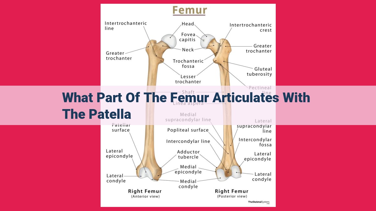
The patella articulates with the anterior surface of the distal femur, specifically the patellar surface. This surface is located between the medial and lateral condyles of the femur and is formed by the trochlea, a groove-like depression. The trochlea guides the patella during knee flexion and extension, allowing for smooth movement and stability of the joint.
Distal End of the Femur
- Discuss the anatomy and location of the distal end of the femur.
- Describe its articular surfaces (medial and lateral condyles) and their roles in articulation.
- Explain the significance of the intercondylar fossa and its role in ligament attachment.
Exploring the Distal End of the Femur: A Deeper Dive
The distal end of the femur, the longest bone in the human body, holds a crucial role in our musculoskeletal system. Let’s delve into its anatomy and significance:
Location and Anatomy
Nestled at the lower end of the thigh bone, the distal femur forms the knee joint with the tibia (shinbone) and patella (kneecap). It’s a bulbous structure with distinctive features that allow for smooth articulation and stability:
- Condyles: The condyles are two prominent projections on either side of the distal femur. The medial condyle faces inward towards the body, while the lateral condyle faces outward.
- Intercondylar Fossa: This deep depression between the condyles serves as an attachment point for ligaments, providing stability to the knee joint.
- Intercondylar Eminence: A small elevation within the intercondylar fossa, it further enhances ligament attachment.
Articular Surfaces
The medial and lateral condyles form the articular surfaces of the distal femur. These surfaces are covered in cartilage and interact with the bones of the lower leg:
- Medial Condyle: It articulates with the medial condyle of the tibia.
- Lateral Condyle: It articulates with both the lateral condyle of the tibia and the head of the fibula.
Significance of the Distal Femur
The distal end of the femur plays a crucial role in knee function:
- Weight Bearing: The condyles bear the weight of the body during standing and walking, distributing forces evenly across the joint.
- Movement: The smooth articulation between the femur and tibia allows for flexion (bending) and extension (straightening) of the knee.
- Ligament Attachment: The intercondylar fossa provides a secure attachment point for the crucial ligaments of the knee, including the anterior cruciate ligament (ACL) and posterior cruciate ligament (PCL), helping to prevent joint instability.
The Patellar **Surface of the **Femur: A Closer Look**
The distal end of the femur, the large bone in the upper leg, comprises the patellar surface, an integral component of the knee joint. This smooth, oval-shaped surface faces anteriorly and articulates directly with the patella (kneecap). The patellar surface is a vital component in the mechanics of knee flexion and extension.
Nestled within the patellar surface is the trochlea, a groove that facilitates the gliding movement of the patella. This intricate structure ensures that the patella remains in place during knee motion, preventing its dislocation.
The patellofemoral joint, formed by the patella’s articulation with the patellar surface of the femur, plays a crucial role in transmitting forces across the knee. The patella acts as a lever, increasing the efficiency of quadriceps muscle action during knee extension.
Understanding the anatomy and biomechanics of the patellar surface is essential for healthcare professionals to diagnose and treat knee injuries effectively. By delving into its intricate details, we can unravel the secrets of this fascinating joint and appreciate its vital role in human movement.
