Neurotransmitter Vesicles: Orchestrating Neuronal Communication

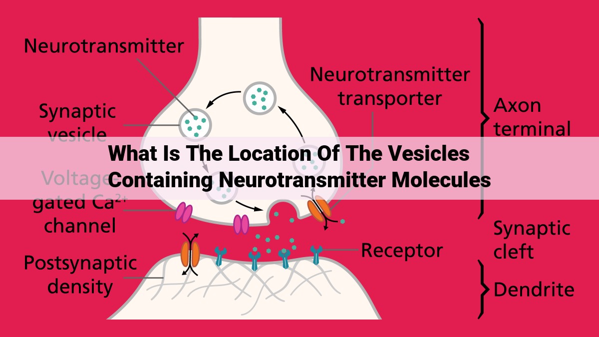
The vesicles containing neurotransmitter molecules are located at the presynaptic membrane, storing and releasing them into the synaptic cleft. Dendritic vesicles, located in dendrites, receive and process signals. Myelinated axons contain vesicles within the myelin sheath, protected by oligodendrocytes for efficient signal transmission. Unmyelinated axons also have vesicles, with axon terminals and Schwann cells aiding vesicle transport and signaling. These diverse locations enable vesicles to orchestrate neuronal communication, facilitating the storage, release, reception, and processing of neurotransmitters.
Synaptic Vesicles: The Storage and Release Powerhouses of Neuronal Communication
In the bustling metropolis of the neuron, there exists a critical component that orchestrates the vital exchange of information: synaptic vesicles. These tiny, sac-like structures are the guardians of neurotransmitters, the chemical messengers that allow neurons to communicate.
Residing at the presynaptic membrane, these vesicles act as storage depots for neurotransmitters. When an electrical signal, known as an action potential, reaches the presynaptic membrane, calcium channels open, causing an influx of calcium ions. This influx triggers the fusion of synaptic vesicles with the membrane, releasing their neurotransmitter cargo into the synaptic cleft, the narrow gap between neurons.
Once released, neurotransmitters traverse the synaptic cleft to bind to receptors on the postsynaptic membrane of the receiving neuron. This binding initiates a cascade of events that can either excite or inhibit the postsynaptic neuron, allowing for the transmission of signals throughout the nervous system.
Vesicles in Dendrites: Receiving and Processing Signals
- Describe the location and function of vesicles in dendrites.
- Explain the importance of postsynaptic membrane, postsynaptic density, and their interaction with dendritic vesicles.
Vesicles in Dendrites: The Unsung Heroes of Neural Communication
In the intricate world of neurons, vesicles serve as tiny but indispensable messengers. While often overshadowed by their counterparts in axons, dendritic vesicles play a crucial role in the reception and processing of signals.
Location and Function
Dendritic vesicles reside in the dendrites, the intricate branches that emanate from the neuron’s cell body. They are responsible for receiving signals from other neurons through neurotransmitters, chemical messengers that bind to specific receptors on the dendrite.
Postsynaptic Membrane and Density
Upon reaching the dendrite, neurotransmitters bind to receptors on the postsynaptic membrane, the membrane that lines the dendritic shaft. This binding triggers a cascade of events, including the opening of ion channels that allow charged ions to flow in or out of the neuron.
The postsynaptic density (PSD) is a specialized area of the postsynaptic membrane where most neurotransmitter receptors are concentrated. The PSD also contains scaffolding proteins that anchor receptors and other molecules involved in signal transduction.
Vesicles in Action
Dendritic vesicles are filled with reuptake transporters, proteins that bind to and remove neurotransmitters from the synaptic cleft, the narrow gap between the presynaptic and postsynaptic neurons. By removing neurotransmitters, these vesicles prevent overstimulation of the neuron and allow for precise control of signal termination.
In addition to their reuptake function, dendritic vesicles also play a role in retrograde signaling. When neurotransmitters bind to receptors on the postsynaptic membrane, they can trigger signals that travel back to the presynaptic neuron, modulating its activity. Dendritic vesicles are involved in this retrograde signaling process, transporting molecules that can influence neurotransmitter release from the presynaptic terminal.
Often overlooked but vital to the symphony of neural communication, dendritic vesicles are the unsung heroes of signal reception and processing. They ensure precise and controlled signaling, allowing neurons to adapt to changing inputs and engage in complex cognitive processes.
Vesicles in Myelinated Axons: The Guardians of Expeditious Communication
Vesicles, the tiny cellular compartments, play a vital role in the intricate dance of neuronal communication. Their presence in myelinated axons grants a protective sanctuary and fuels efficient signal transmission.
Nestled within the myelin sheath, these vesicles have a pivotal mission: safeguarding the delicate cargo of electrical impulses. The myelin sheath, a multi-layered insulating blanket formed by oligodendrocytes, wraps around the axon, creating a protective barrier against external interference. This insulative armor ensures the safe and rapid conduction of electrical signals, preventing dissipation and maintaining signal integrity.
Oligodendrocytes, the master architects of myelination, not only provide protection but also actively participate in the vesicle dance. They extend their myelinating processes, wrapping around the axon like an elaborate dance partner, ensuring the smooth and efficient relay of electrical impulses. The interaction between vesicles and myelin sheath in myelinated axons is a symphony of precision, enabling neurons to communicate swiftly and reliably.
Vesicles in Unmyelinated Axons: Maintaining Communication
In the bustling world of neuronal communication, vesicles serve as the unsung heroes, orchestrating the flow of vital information along unmyelinated axons. These slender, unencapsulated fibers are the primary channels through which neurons send signals to their distant targets. And within these axons, vesicles perform a critical mission: transporting neurotransmitters and facilitating their release.
Vesicles in unmyelinated axons are located in close proximity to the axon terminal, the specialized region where signals are transmitted. These vesicles contain a host of neurotransmitters, which are the chemical messengers that neurons use to communicate with each other. When an electrical signal reaches the axon terminal, it triggers the release of vesicles, unleashing a surge of neurotransmitters into the synaptic cleft, the narrow gap between the presynaptic and postsynaptic neurons.
Crucial to this process is the interplay between vesicles and Schwann cells, the glial cells that envelop unmyelinated axons. Schwann cells provide structural support to the axon and regulate the transport of vesicles along its length. They secrete a variety of molecules that guide and protect vesicles as they navigate the axon’s intricate terrain towards the axon terminal, ensuring their timely and accurate delivery.
Once vesicles reach the axon terminal, they undergo exocytosis, the process of fusing with the presynaptic membrane and releasing their neurotransmitter cargo into the synaptic cleft. These neurotransmitters then bind to receptors on the postsynaptic membrane, triggering a cascade of events that either excite or inhibit the postsynaptic neuron, relaying the message further along the neural pathway.
In summary, vesicles in unmyelinated axons play a pivotal role in maintaining communication between neurons. Their ability to store, transport, and release neurotransmitters enables the efficient and targeted transmission of signals, orchestrating the intricate symphony of neural communication that underlies our thoughts, actions, and experiences.