Mitotic Index: A Vital Measure Of Cell Proliferation For Cancer Diagnosis And Tissue Engineering
Mitotic index, a measure of cell proliferation, indicates the percentage of cells actively dividing. Calculated by counting mitotic figures (cells in mitosis) among total cells, it reflects the growth and proliferation rate of a cell population. This index plays a crucial role in cancer diagnosis, prognosis, and tissue engineering. By assessing the mitotic index, researchers and clinicians gain insights into tumor aggressiveness, tissue regeneration, and cellular growth dynamics.
The Mitotic Index: A Tale of Cell Proliferation
In the bustling metropolis of life’s processes, cells play a pivotal role like tireless workers. Understanding their growth and division is crucial to unravelling the secrets of life’s enigmatic dance. One tool that provides invaluable insights into this cellular ballet is the enigmatic concept of the mitotic index.
The mitotic index is a measure that eloquently captures the pace at which cells are proliferating or dividing. It’s a window into the cellular machinery that governs life’s most fundamental processes like growth, repair, and renewal. By studying the mitotic index, scientists can unravel the secrets of cellular behavior and pave the way for new discoveries in medicine and biology.
Understanding Mitotic Figures and the Cell Cycle: A Journey into Cellular Division
In the realm of cell biology, the mitotic index holds a profound significance in unraveling the mysteries of cell growth and reproduction. To comprehend the essence of this vital metric, it’s imperative to delve into the fascinating world of mitotic figures and the intricate dance of the cell cycle.
Mitotic Figures: The Footprints of Cell Division
Imagine a cell preparing to divide. Within its cytoplasm, threadlike structures known as chromosomes start to condense and become visible under a microscope. At this stage, the cell has entered the realm of mitosis, a highly orchestrated process that ensures the faithful distribution of genetic material into two daughter cells. As mitosis progresses, the chromosomes undergo a series of dynamic transformations, creating distinct stages that can be observed as mitotic figures.
Prophase: The Stage of Preparation
Like a symphony about to unfold, prophase marks the transition from the resting state of interphase to active cell division. The chromatin, a complex of DNA and proteins, condenses into visible chromosomes, each carrying two copies (sister chromatids) of the genetic information. The nuclear envelope, which encloses the nucleus, begins to break down.
Metaphase: The Line-Up
Metaphase is the stage of precision. The chromosomes align themselves at the equator of the cell, forming a flat plate called the metaphase plate. Each chromosome is attached to spindle fibers, microtubule structures that will guide the chromosomes during cell division. These fibers exert pulling forces, ensuring proper alignment and separation of sister chromatids.
Anaphase: The Separation
Tension builds in anaphase as the spindle fibers shorten, pulling the sister chromatids apart. Each chromatid, now an independent chromosome, migrates towards opposite poles of the cell. As the chromosomes continue to move, the cell elongates, preparing for the final stage of mitosis.
Telophase: The Restoration
In telophase, two new nuclear envelopes form around the two sets of chromosomes. The spindle fibers disintegrate, and the chromatin gradually decondenses, returning to its resting state. The cytoplasm divides into two, a process called cytokinesis, giving rise to two genetically identical daughter cells.
Interphase: The Rest and Preparation Zone
After completing a round of mitosis, cells enter interphase, a period of intense growth and preparation for the next division. Interphase consists of three subphases:
-
G1 phase: The cell increases in size and synthesizes proteins and other molecules necessary for growth.
-
S phase: The DNA in the cell is replicated, creating two identical copies of each chromosome.
-
G2 phase: The cell makes final preparations for mitosis, producing spindle fibers and other proteins needed for cell division.
The ability to accurately identify mitotic figures and understand the stages of the cell cycle is essential for researchers and clinicians. From uncovering the mysteries of cell growth to diagnosing diseases, the mitotic index remains a powerful tool in the exploration of life’s fundamental processes.
Calculating Mitotic Index: A Measure of Cellular Proliferation
Determining the rate at which cells divide is crucial for understanding cell growth and proliferation. The mitotic index, a valuable tool in this regard, offers insights into the dynamics of cell division.
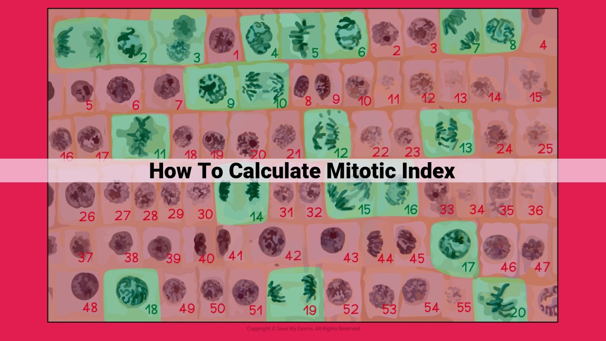
Calculating Mitotic Index:
The mitotic index is calculated using the formula:
Mitotic Index = Number of Mitotic Figures / Total Number of Cells
Mitotic figures refer to cells undergoing mitosis, the process of cell division.
Steps for Conducting a Mitotic Index Count:
- Tissue Preparation: Tissue samples are collected and prepared for microscopic examination by staining to visualize mitotic figures.
- Counting Mitotic Figures: The number of mitotic figures is counted under a microscope.
- Total Cell Count: The total number of cells in the sample is determined by counting all cells within a specific area.
- Calculation: The mitotic index is calculated using the formula provided above.
Significance of Mitotic Index:
The mitotic index serves as a quantitative measure of cell proliferation. A high mitotic index indicates a rapid rate of cell division, often associated with growth, repair, or pathological conditions such as tumors. Conversely, a low mitotic index suggests a slow or dormant state of cell proliferation.
Monitoring mitotic index over time allows researchers and clinicians to assess changes in cell proliferation, providing valuable insights into cell cycle dynamics, growth patterns, and disease progression. It aids in diagnosing and prognosing certain diseases, including cancer.
Applications of Mitotic Index
Cancer Diagnosis and Prognosis
In the realm of oncology, mitotic index plays a pivotal role in unraveling the mysteries of cancer. By meticulously calculating the number of cells dividing within a tumor sample, pathologists can discern the growth rate and aggressiveness of the malignancy. Tumors with a high mitotic index indicate a more proliferative nature, suggesting a faster-growing and potentially more dangerous disease. This valuable information aids in determining the appropriate treatment strategies and predicting patient outcomes.
Tissue Engineering and Regenerative Medicine
Beyond cancer diagnosis, mitotic index finds its application in the burgeoning field of tissue engineering and regenerative medicine. As scientists strive to repair damaged tissues and organs, they rely on mitotic index to monitor and manipulate cell growth. By carefully controlling the mitotic rate, researchers can guide the formation of new tissues, paving the way for transformative treatments for a wide range of conditions.
The mitotic index serves as an indispensable tool in both medical diagnostics and regenerative medicine. Its ability to quantify cell proliferation empowers researchers and clinicians to better understand the intricate workings of the human body, both in health and disease. As our scientific understanding continues to evolve, the applications of mitotic index are bound to expand, unlocking new possibilities for healing and innovation.
