Unveiling The Critical Preparations For Mitosis: A Comprehensive Guide
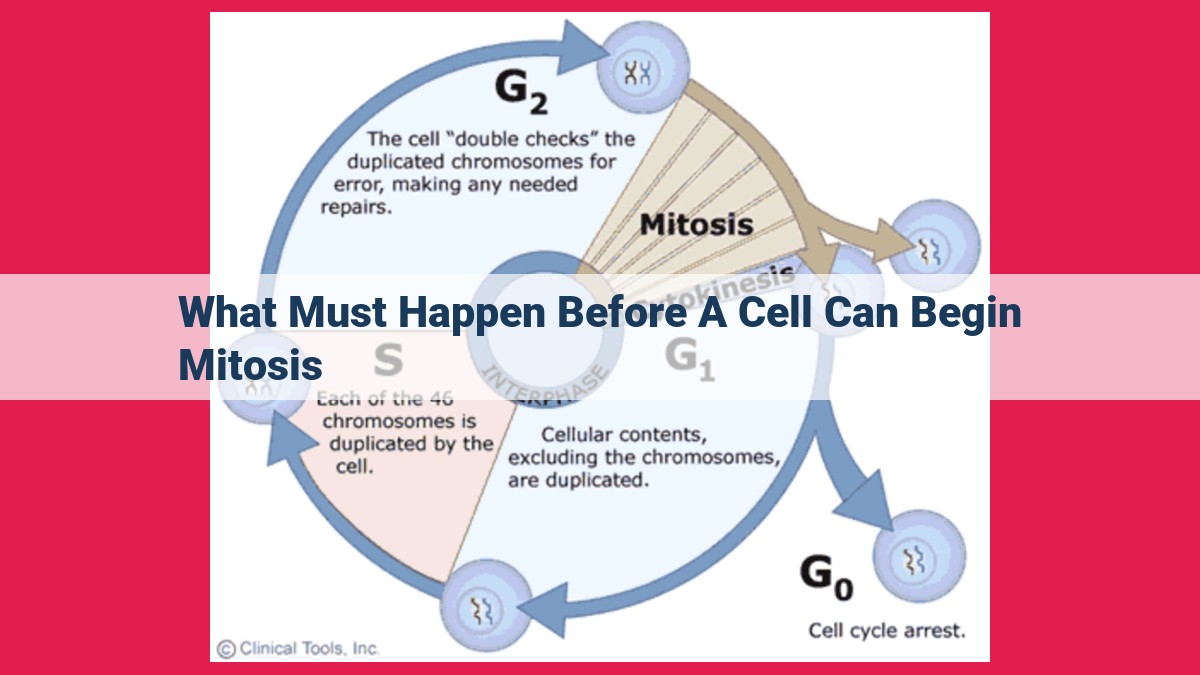
Before mitosis, cells undergo several critical preparations: DNA replication duplicates chromosomes, ensuring accurate genetic inheritance; the nuclear envelope disintegrates, granting access to chromosomes; the mitotic spindle assembles as a framework for chromosome separation; and centrioles migrate to opposing poles of the cell, forming poles and organizing microtubules. These events lay the groundwork for the precise division of genetic material during mitosis.
Chromosome Duplication: The Foundation of Genetic Inheritance
In the intricate tapestry of life, cells hold the blueprint for our existence, carrying the genetic information that governs every aspect of our being. For life to propagate, cells must meticulously divide, ensuring each daughter cell receives an exact copy of the parent cell’s genetic material. At the heart of this remarkable process is chromosome duplication, a fundamental event that lays the foundation for accurate genetic inheritance.
The chromosomes within our cells are thread-like structures composed of DNA, the molecule that stores genetic information. During chromosome duplication, each chromosome is meticulously replicated, producing two identical copies known as sister chromatids. This process relies on DNA replication, a complex and precise molecular dance in which the DNA double helix unwinds and each strand serves as a template for the synthesis of a new complementary strand.
This intricate duplication process ensures that each daughter cell receives a complete and accurate set of genetic instructions, preserving the genetic integrity of our species. It is a testament to the remarkable precision and complexity of cellular processes, a symphony of molecular events that enables the continuity of life.
Nuclear Membrane Breakdown: Breaking Down Barriers for Mitosis
The nuclear membrane, also known as the nuclear envelope, is a double-membrane structure that encloses the cell’s genetic material, DNA. It plays a crucial role in regulating the entry and exit of molecules into and out of the nucleus. But during mitosis, the process of cell division, the nuclear membrane undergoes a remarkable metamorphosis.
Disassembly of the Nuclear Envelope
As the cell prepares for mitosis, the nuclear membrane begins to disassemble, allowing access to the chromosomes that are coiled within. This process is triggered by a group of proteins called nuclear pore complexes (NPCs). NPCs span both nuclear membranes and create channels for the exchange of molecules between the nucleus and the cytoplasm.
During disassembly, the NPCs detach from the nuclear membrane, and the two membranes fuse together, forming a single continuous membrane. This fusion further weakens the nuclear envelope and allows it to break down completely.
Role of the Nuclear Lamina
Supporting the nuclear membrane from the inside is a meshwork of proteins called the nuclear lamina. The nuclear lamina provides structural support to the nucleus and helps maintain its shape. During mitosis, the nuclear lamina undergoes a series of modifications that contribute to the breakdown of the nuclear envelope.
As the cell enters mitosis, the nuclear lamina becomes phosphorylated, or modified with phosphate groups. This phosphorylation weakens the interactions between the nuclear lamina and the nuclear membrane, facilitating the disassembly of the envelope. Once the nuclear membrane is completely broken down, the nuclear lamina disperses throughout the cytoplasm.
Significance of Nuclear Envelope Breakdown
The breakdown of the nuclear membrane is a crucial step in mitosis. It allows the mitotic spindle, a structure made of microtubules, to enter the nucleus and access the chromosomes. The mitotic spindle is responsible for separating the chromosomes during cell division, ensuring that each daughter cell receives a complete set of genetic material.
Without the breakdown of the nuclear membrane, the mitotic spindle would not be able to reach the chromosomes, and cell division would not be possible. Therefore, the disassembly of the nuclear envelope is an essential step in the intricate process of mitosis, ensuring the accurate and equitable distribution of genetic material to daughter cells.
Mitotic Spindle Formation: The Framework for Chromosome Separation
As mitosis unfolds, a remarkable structure emerges within the cell, guiding the separation of chromosomes: the mitotic spindle. This intricate latticework serves as the scaffold upon which chromosomes align and divide.
The mitotic spindle is composed of microtubules, tiny cylindrical structures made up of tubulin proteins. These microtubules emanate from opposite ends of the cell, known as the spindle poles. Each spindle pole contains a structure called a centrosome, which harbors a pair of centrioles.
Centrioles are short, cylindrical structures that function as microtubule-organizing centers. During mitosis, they migrate to opposite poles of the cell, establishing the anchors for spindle microtubules. These microtubules extend out from the centrosomes, forming long fibers called spindle fibers.
The spindle fibers play a crucial role in chromosome separation. They attach to the kinetochores, specialized structures on the chromosomes. As the spindle fibers shorten, they pull the chromosomes towards the spindle poles, ensuring their equitable distribution into daughter cells.
The spindle poles serve as organizing hubs for microtubule assembly. They contain proteins that promote microtubule growth and crosslinking, creating the rigid structure necessary for chromosome segregation. The spindle poles also undergo dynamic movements during mitosis, ensuring proper chromosome alignment and separation.
Thus, the mitotic spindle, with its intricate array of microtubules, centrioles, and spindle fibers, provides the architectural framework for chromosome separation during mitosis. This process is vital for ensuring the faithful inheritance of genetic material in all dividing cells.
Centriole Movement: Establishing Poles of Division
Before a cell can embark on the complex journey of mitosis, it meticulously orchestrates a series of events to ensure an accurate and successful cell division. Among these critical steps is the movement of centrioles, tiny cylindrical structures that play a pivotal role in establishing the poles of division.
Location and Function of Centrioles
Centrioles reside within the cell’s centrosome, a small, but mighty organelle that serves as the organizational and microtubule-nucleating hub. These microtubules, the building blocks of the mitotic spindle, radiate outwards from the centrioles like the spokes of a wheel, providing the framework for chromosome separation.
Migration to Opposite Poles
As the cell prepares for mitosis, the centrosomes, housing the centrioles, duplicate themselves and migrate to opposite ends of the cell. This migration is orchestrated by motor proteins that pull the centrosomes along microtubule tracks. The formation of two distinct poles is essential for organizing the mitotic spindle and ensuring the equal distribution of chromosomes to daughter cells.
Pole Bodies: Guiding Forces
Once the centrosomes reach the poles, they mature into specialized structures known as pole bodies. These complex structures are comprised of numerous smaller substructures that serve as docking stations for microtubules. By organizing the attachment of microtubules, the pole bodies define the spindle poles and guide the formation of the mitotic spindle.
The precise movement of centrioles to opposite poles is a critical step in ensuring the success of mitosis. The poles serve as anchors for the mitotic spindle, enabling the equal distribution of chromosomes to daughter cells. This elaborate choreography highlights the complexity and regulation of mitosis, a process that underlies growth, development, and the very essence of life itself.
