Microfilaments: Key Players In Cell Division

Microfilaments, composed of actin proteins, play a vital role in cell division, particularly during cytokinesis. They form a contractile ring through assembly and disassembly processes, constricting the cell membrane and facilitating the separation of daughter cells. Microfilaments interact with motor proteins like myosin to generate force and maintain cell shape. Regulatory mechanisms involving signaling pathways and post-translational modifications control microfilament dynamics, ensuring accurate cell division.
The Unsung Heroes of Cell Division: Microfilaments
In the realm of cellular biology, cell division stands as a pivotal process that ensures the growth, development, and renewal of life. At the heart of this intricate dance lies a network of microscopic filaments known as microfilaments, playing a crucial role in orchestrating the meticulous separation of one cell into two.
Microfilaments: The Building Blocks of Cell Division
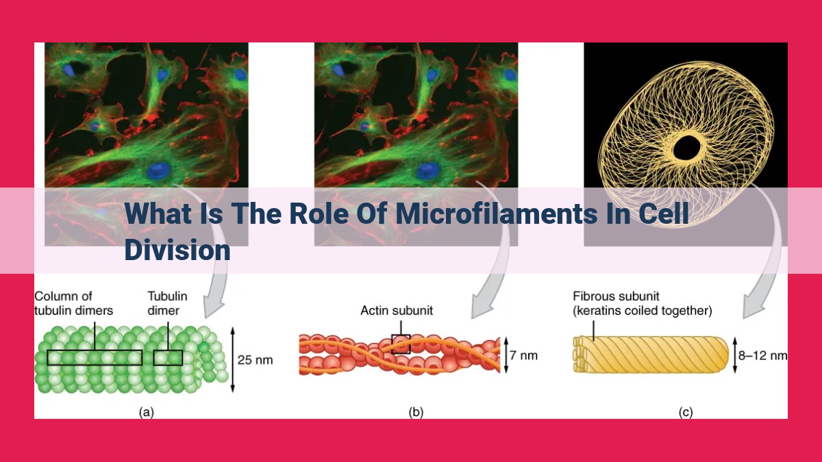
Picture microfilaments as slender, thread-like structures that form a dynamic scaffold within cells. Composed primarily of the protein actin, these filaments possess unique properties that make them ideally suited for their role in cell division. They showcase remarkable flexibility, allowing them to bend and reshape with ease, and exhibit polarity, meaning they have distinct ends with different chemical compositions. Moreover, microfilaments have an intrinsic ability to polymerize and depolymerize, a process that allows them to assemble and disassemble rapidly in response to cellular cues.
The Invisible Forces: Microfilaments in Cytokinesis
During the intricate process of cytokinesis, the final stage of cell division, microfilaments take center stage. They orchestrate a symphony of events that ensures the equal partitioning of cellular contents into two daughter cells.
Contraction and Separation: The Contractile Ring Formation
As cytokinesis unfolds, microfilaments gather in a specific region of the cell, forming a contractile ring. This assembly process is driven by the polymerization of actin filaments, facilitated by the action of motor proteins that act as molecular bridges. The contractile ring encircles the cell’s midpoint, resembling a belt tightening around its equator.
With the contractile ring in place, the show begins. Motor proteins like myosin step into action, harnessing their ability to walk along the microfilaments. As they do so, they drag adjacent filaments closer together, causing the contractile ring to constrict.
The Disassembly Dance: Completion of Cytokinesis
As the contractile ring tightens, a wave of microfilament disassembly sweeps across the ring. This disassembly is orchestrated by a protein complex known as the actomyosin disassembly machinery. This machinery snips apart the polymers of actin, allowing the contractile ring to disengage and separating the two daughter cells.
A Helping Hand: Interactions with Other Players
Microfilaments do not work in isolation; they collaborate with a cast of cellular components to ensure the smooth execution of cell division.
Motor Proteins: The Muscle of Microfilaments
Motor proteins stand out as crucial partners of microfilaments. Myosin, the flagship motor protein, binds to actin filaments and uses its molecular feet to walk alongside them. This movement drives the contraction of the microfilament network, essential for cytokinesis.
Cell Shape: Maintaining Harmony
In addition to their role in cytokinesis, microfilaments also play a vital role in maintaining cell shape. They form a framework that gives cells their structure and allows them to change shape as needed.
Regulation: Orchestrating the Microtubule Symphony
The dynamics of microfilaments are tightly regulated by a complex interplay of signaling pathways and post-translational modifications. These regulatory mechanisms ensure that microfilaments are assembled, disassembled, and reorganized at the right time and place to facilitate cell division.
Microfilaments are not just bystanders in cell division; they are the unsung heroes, the driving force behind the precise and efficient separation of cells. Their dynamic behavior, orchestrated by a symphony of interactions and regulations, ensures the successful completion of this fundamental process that underpins the very essence of life.
By understanding the intricate workings of microfilaments, we gain a deeper appreciation for the incredible complexity and elegance of cellular life. Their role in cell division is a testament to the remarkable dance that cells perform, ensuring the growth, development, and renewal of all living organisms.
Understanding Microfilaments
- Describe the structure, properties, polarity, and polymerization dynamics of microfilaments.
Understanding Microfilaments
Micofilaments are the unsung heroes of cell division, the process that ensures the growth and renewal of life itself. These tiny cellular structures play a crucial role in shaping and dividing cells, making them one of the key players in the intricate dance of life.
At their core, microfilaments are thin, flexible rods made of a protein called actin. They resemble tiny threads that crisscross the cell, providing structural support and flexibility. These threads, only about six nanometers in diameter, are often overlooked but are surprisingly strong and dynamic.
The polarity of microfilaments is a fascinating aspect of their structure. Each filament has two ends, a plus end and a minus end. The plus end is the growing end, where new actin subunits are added, while the minus end is the disassembling end. This polarity allows microfilaments to assemble and disassemble in a controlled manner, a process essential for their diverse functions.
The polymerization dynamics of microfilaments are remarkable. They can polymerize and depolymerize rapidly, changing their length and shape in response to cellular cues. This dynamic behavior enables microfilaments to reshape the cell and facilitate processes such as cell division, movement, and adhesion.
Microfilaments: The Unsung Heroes of Cell Division
Cell division, a fundamental process in life, is a meticulously choreographed dance of cellular components, with microfilaments playing a pivotal role as the “muscle fibers” of the division machinery. These tiny protein filaments, composed of actin monomers, are not only crucial for cell shape and movement but also for a process called cytokinesis.
Cytokinesis: The Final Cut
Cytokinesis is the last and arguably most critical stage of cell division, where the dividing cell splits into two distinct daughter cells. This complex process relies heavily on microfilaments to ensure an equitable distribution of cellular components.
Contractile Ring Assembly: A Molecular Tug-of-War
The first step of cytokinesis involves the assembly of a contractile ring made of microfilaments. This ring forms at the equator of the dividing cell and acts like a molecular belt, constricting and pinching the cell membrane. The assembly process is driven by motor proteins, such as myosin, which interact with microfilaments and use ATP to “walk” along them, causing them to slide past each other and tighten the ring.
Contractile Ring Disassembly: A Delicate Balance
Once the contractile ring has completed its constriction, it must be disassembled to allow the daughter cells to separate. This disassembly is a finely tuned process that involves the cleavage of actin filaments by enzymes called actin depolymerizing factors. By breaking down the microfilament scaffold, these enzymes allow the daughter cells to pull apart cleanly.
Interplay with Related Components: A Collaborative Effort
Microfilaments do not work in isolation during cytokinesis. They interact with a host of other cellular components to ensure a successful division:
- Motor proteins, like myosin, provide the force necessary for microfilament movement and ring constriction.
- Other cytoskeletal components, such as microtubules, help maintain the cell’s overall shape and guide the division furrow.
Regulation of Microfilament Dynamics: A Signaling Symphony
The dynamics of microfilament assembly and disassembly during cytokinesis are tightly regulated by a complex symphony of signaling pathways. Key players in this orchestra include:
- Rho GTPases, small proteins that control the activity of various actin-regulatory proteins.
- Rho-associated kinases (ROCK), which promote microfilament assembly and contraction.
- Post-translational modifications, such as phosphorylation, which can alter microfilament behavior and localization.
Without microfilaments, cell division would be a chaotic free-for-all, with cells unable to divide properly or distribute their contents equally. Their ability to form dynamic structures and interact with other cellular components makes them indispensable for this fundamental biological process. Understanding the intricate workings of microfilaments in cytokinesis thus provides invaluable insights into the complexities of cell biology and the very essence of life itself.
Interactions with Related Components
Microfilaments don’t work in isolation. They collaborate with various cellular components to orchestrate the intricate process of cell division.
A. Motor Proteins: A Dynamic Partnership
Among the most crucial partnerships is that with motor proteins, especially myosin. These molecular workhorses use energy to “walk” along microfilaments. Acting like tiny motors, myosin molecules pull microfilaments towards each other, generating the force necessary for cell division.
B. Cell Shape: A Symphony of Forces
Microfilaments play a pivotal role in maintaining cell shape during division. By interacting with other cytoskeletal components, such as microtubules, they form a dynamic network that shapes the cell. This network allows the cell to maintain its structural integrity while undergoing division.
Together, these interactions between microfilaments and other cellular components orchestrate a complex dance that ensures precise and efficient cell division, a fundamental process for growth, development, and health.
Regulation of Microfilament Dynamics
The intricate dance of microfilaments during cell division is a finely regulated process. Orchestrating this dynamic behavior is a symphony of signaling pathways and post-translational modifications.
Signaling Pathways
Rho GTPases, the masters of microfilament fate, wield their power through a cascade of signaling events. When activated, they recruit and activate Rho-associated kinases (ROCKs), the generals of microfilament assembly. ROCKs, in turn, phosphorylate countless targets, including myosin light chain kinase (MLCK), the key to microfilament contraction.
Post-translational Modifications
Microfilaments are not passive bystanders in this regulatory ballet. Instead, they undergo a series of transformative modifications that dramatically alter their behavior. Phosphorylation, the addition of phosphate groups, is a major player. By adding or removing these chemical tags, cells can fine-tune microfilament stability, assembly, and disassembly.
During cytokinesis, the microfilaments in the contractile ring are subjected to a rigorous barrage of phosphorylation. This molecular makeover initiates their disassembly, signaling the final separation of daughter cells.