Essential Guide To Magnifying Bacteria With Compound Microscopes For Optimal Observation
To observe bacteria, a compound microscope is essential. Objective lenses, ranging from 10x to 100x, determine the magnification level. The eyepiece lens further magnifies the image, with 10x being common. To achieve the optimal magnification for studying bacteria, consider their size and desired detail level. A typical range is between 1000x (10x objective lens x 10x eyepiece lens) to 1500x (15x objective lens x 10x eyepiece lens) to balance resolution and image clarity.
- Dive into the fascinating world of bacteria and their importance in our lives.
Delving into the Microscopic Realm of Bacteria: A Journey of Discovery
In the vast expanse of the microscopic world, bacteria reign supreme. These tiny organisms, invisible to the naked eye, play pivotal roles in our lives and the functioning of our planet. They are essential for nutrient cycling, waste decomposition, and even food production. To delve into this hidden realm, scientists have harnessed the power of magnification, enabling us to unlock the secrets of these microscopic wonders.
Compound Microscopes: Gateway to Bacterial Visualization
The compound microscope is a gateway to the intricate world of bacteria. Its multiple lenses work in tandem to magnify images, allowing us to observe the fine details of these microorganisms. The objective lens, located near the specimen, gathers light and focuses it on the image plane, while the eyepiece lens further magnifies this image for our eyes to examine.
Dissecting Microscopes: Unveiling Macroscopic Microbial Structures
In contrast to compound microscopes, dissecting microscopes provide a lower magnification range. This perspective allows us to study larger bacterial structures and examine the overall morphology of colonies or samples. By using different lenses and techniques, scientists can gain insights into the organization and behavior of bacteria at a macroscopic level.
Magnification: Unlocking Bacterial Details
Magnification is the cornerstone of bacterial observation. By increasing magnification, we can resolve finer details and observe the ultrastructure of bacteria, including their cell walls, membranes, and other internal components. Different magnification levels are suitable for different purposes, and choosing the appropriate level is crucial for optimizing the information obtained.
Objective Lens: Magnification Magnified
The objective lens plays a vital role in determining both magnification and image quality. Different objective lenses have different focal lengths, which affect the level of magnification. By rotating the nosepiece, scientists can switch between objective lenses and seamlessly adjust the magnification to suit their needs.
Eyepiece Lens: Enhancing the Magnified Image
The eyepiece lens is the final component in the magnification chain. It further magnifies the image produced by the objective lens, making it more comfortable for our eyes to observe. Eyepieces typically have a magnification of 10x or 15x, providing a comfortable viewing experience while maintaining image clarity.
Abbe Condenser: Optimizing Illumination for Enhanced Viewing
To achieve optimal image quality, proper illumination is essential. The Abbe condenser, located beneath the stage, focuses light onto the specimen, ensuring even illumination and reducing glare. This enhances contrast and improves the visibility of bacterial structures.
Stage Micrometer: Ensuring Accurate Magnification Calibration
Calibrating the microscope is crucial for accurate measurements and comparisons. A stage micrometer, a slide with etched lines of known dimensions, is used to determine the exact magnification of each objective lens. By observing the micrometer under the microscope, scientists can calculate the magnification and ensure consistent and reliable measurements.
Optimal Magnification for Bacterial Observation
The ideal magnification range for studying bacteria varies depending on the size and complexity of the organism. For general observation, a magnification of 1000x (100x objective and 10x eyepiece) is suitable. However, higher magnifications (e.g., 4000x) may be necessary to reveal intricate details such as flagella or pili.
Compound Microscope: Unveiling the Microscopic Realm of Bacteria
Embark on a captivating journey into the microscopic world of bacteria, where the compound microscope emerges as an indispensable tool for revealing hidden wonders. This magnificent instrument empowers researchers to delve into the intricacies of bacterial structure, unlocking a wealth of knowledge that shapes our understanding of these ubiquitous organisms.
At the heart of a compound microscope lies its intricate assembly of lenses. The objective lens acts as the primary magnifier, drawing in the minute details of bacteria. As light passes through the specimen, the condenser optimizes illumination, enabling clear and vibrant observations. The eyepiece lens further amplifies the image, presenting a magnified spectacle that astounds the viewer.
Magnification, a fundamental concept in microscopy, refers to the ability to enlarge an image. By understanding the interplay between the objective and eyepiece lenses, researchers can tailor the magnification to suit the specific characteristics and desired level of detail of the bacteria being studied. This precise control grants unparalleled insights into the intricate structures and behaviors of these microbial marvels.
The compound microscope has revolutionized bacterial examination, allowing researchers to explore their intricate structures, witness their dynamic interactions, and unravel their vital roles in our ecosystem. As we peer through the lens, we uncover a hidden world teeming with life, where bacteria play a profound role in shaping our planet and our own biology.
Dissecting Microscope: Unraveling the Macroscopic Realm of Microbial Structures
In the intricate tapestry of life, the world of bacteria beckons us to explore the microscopic wonders that shape our existence. While compound microscopes grant us a glimpse into the cellular complexities of these tiny organisms, dissecting microscopes offer a unique perspective, unveiling the macroscopic structures that contribute to their distinct forms and functions.
Unlike their compound counterparts, dissecting microscopes utilize lower magnification and a stereoscopic view to provide a three-dimensional image of the specimen. This allows researchers to observe the overall morphology, texture, and coloration of bacterial colonies, as well as dissect and manipulate them without disrupting their delicate structures.
Dissecting microscopes come equipped with binocular lenses and a base plate with a stage for holding the specimen. The objective lens is typically mounted on an adjustable arm, allowing for precise focus and magnification. Illumination is provided from above and below to ensure optimal visibility.
While compound microscopes excel in revealing the intricate cellular machinery of bacteria, dissecting microscopes are indispensable for studying their larger-scale features. For instance, researchers can examine the biofilms formed by bacterial communities, the flagella that enable their movement, and the spores that allow them to survive harsh conditions.
Dissecting microscopes have also found applications in medical diagnostics, enabling the visualization of bacterial colonies on culture plates and the identification of their unique morphologies. By providing a broader perspective on bacterial organization, dissecting microscopes complement compound microscopes, empowering researchers to delve deeper into the fascinating world of these microscopic marvels.
Magnification: Unraveling the Microscopic World of Bacteria
Delve into the fascinating realm of microbiology, where the microscopic world of bacteria holds profound significance. Unlocking the secrets of these tiny organisms requires specialized tools like microscopes, which are indispensable for unraveling their intricate details. One crucial element in microscopic exploration is magnification, which empowers scientists to magnify the tiniest structures, enabling a profound understanding of bacterial biology.
Magnification: Unveiling the Hidden Details
Magnification is the ability to enlarge the apparent size of an object, revealing details that would otherwise remain concealed. In the context of bacterial observation, the level of magnification directly impacts the clarity and resolution of the image, allowing researchers to discern finer details and morphological characteristics.
Optimal Magnification for Bacterial Observation
The choice of magnification level depends on the specific characteristics of the bacteria being studied. Generally, higher magnification provides greater detail, but it may also reduce the field of view and make it challenging to observe the overall context. Conversely, lower magnification offers a broader perspective, but sacrifices some detail.
For most bacterial observations, a magnification range between 400x and 1000x is ideal. This range allows scientists to observe cellular structures, flagella, and other fine details with sufficient clarity while maintaining a reasonable field of view.
The Impact of Magnification on Bacterial Observation
Magnification not only enhances the visibility of bacterial structures but also influences the accuracy of measurements and interpretations. Accurate magnification calibration is essential to ensure reliable data. This can be achieved using a stage micrometer, a calibrated slide that provides a precise reference for determining the magnification level.
Empowering Researchers with Enhanced Observation
Magnification has revolutionized microbiology, empowering researchers to explore the microscopic world with unprecedented precision. Through the lens of a microscope, scientists can decipher the intricate workings of bacteria, unlocking insights into their behavior, pathogenicity, and potential applications in biotechnology and medicine.
Magnification is a cornerstone of microbial exploration, enabling researchers to delve into the hidden world of bacteria and uncover their secrets. By understanding the principles of magnification and choosing the appropriate level, scientists can optimize their observations and gain a deeper understanding of these fascinating microorganisms.
The Objective Lens: The Magnification Multiplier
In the realm of microbiology, magnification is the key that unlocks the hidden world of bacteria. And at the heart of this magnification power lies the objective lens. This tiny, yet crucial component plays an indispensable role in determining the quality and level of magnification you can achieve when examining your bacterial specimens.
The objective lens is situated at the lower end of the microscope, closest to the specimen. It is composed of multiple glass elements, each carefully designed to bend and focus light in a way that amplifies the image of the specimen. The numerical aperture (NA) of the objective lens, which is a measure of its light-gathering ability, is a key factor in determining the resolution and clarity of the image you obtain.
Each objective lens bears a specific magnification power, ranging from 4X to 100X or even higher. The higher the magnification power, the larger the specimen will appear in the field of view. However, it’s important to note that higher magnification does not always equate to better image quality.
The working distance, which is the space between the objective lens and the specimen, also plays a role in choosing the right objective lens. Longer working distances are ideal for thicker specimens, while shorter working distances provide higher magnification but reduce the space available for manipulating the specimen.
By carefully selecting the appropriate objective lens based on its magnification power, NA, and working distance, you can optimize the visualization of your bacterial specimens and unlock a deeper understanding of their intricate structures and characteristics.
Eyepiece Lens: Unveiling the Microscopic Realm
The eyepiece lens plays a pivotal role in microscopy, magnifying the image further after it has passed through the objective lens. This crucial component enhances the clarity and detail of the magnified specimen, enabling researchers to delve deeper into the microscopic world.
Magnification and Clarity
The eyepiece lens consists of multiple lenses that work in conjunction to enlarge the image produced by the objective lens. By magnifying the image, the eyepiece lens makes it easier to distinguish fine details and structures within the specimen. Additionally, it enhances image clarity, reducing any distortions or aberrations that may have been introduced by the objective lens.
Ergonomics and Viewing Angle
The eyepiece lens also plays a key role in ergonomics and viewing comfort. The diameter of the eyepiece lens affects the field of view, and a larger diameter provides a wider viewing area. Furthermore, the eye relief of the eyepiece lens determines how far the user’s eye can be from the lens while still seeing the full field of view. A longer eye relief provides greater comfort during extended viewing sessions.
Types of Eyepiece Lenses
There are various types of eyepiece lenses, each designed for specific applications. Wide-field eyepieces provide a larger field of view, making them suitable for scanning specimens or observing large areas. High-eyepoint eyepieces offer increased eye relief, reducing eye strain and fatigue. Graduated eyepieces feature a scale or graticule that allows for precise measurements of the specimen.
Calibration and Magnification
To ensure accurate measurements, the eyepiece lens must be calibrated in conjunction with the objective lens. This calibration process involves using a stage micrometer, which is a slide with a known scale of etched lines. By observing the stage micrometer through the microscope, the magnification of the eyepiece lens can be determined and recorded.
The eyepiece lens is an essential component of any microscope, enabling researchers to magnify and enhance the clarity of microscopic specimens. By choosing the appropriate eyepiece lens based on the desired magnification, field of view, and ergonomics, microscopists can optimize their observations and uncover the intricate details of the microscopic world.
The Abbe Condenser: A Guiding Light for Enhanced Bacterial Visualization
In the realm of microbiology, where the microscopic world unfolds its wonders, the Abbe condenser plays a pivotal role in illuminating the intricate details of bacteria. This unsung hero of the microscope optimizes illumination, casting a brighter light on the bacterial realm, enabling researchers to witness their fascinating characteristics with unrivaled clarity.
The Abbe condenser is a lens system positioned beneath the stage of the microscope, between the light source and the specimen. Its primary function is to gather and concentrate light, directing it evenly onto the bacterial sample. Without this condenser, the light would be scattered and unevenly distributed, resulting in a hazy and poorly illuminated image.
The importance of optimal illumination cannot be overstated. Bacteria are typically transparent, making it challenging to distinguish their structures under low light conditions. By enhancing illumination, the Abbe condenser increases the contrast between the bacteria and their surroundings, making them more visible and easier to study.
Furthermore, the condenser also reduces glare and stray light, which can interfere with the image quality. By directing a focused beam of light onto the sample, the condenser helps to eliminate background noise, resulting in a sharper and more detailed image.
In essence, the Abbe condenser is the guiding light for bacterial visualization. It illuminates the microscopic realm, revealing the intricate details that would otherwise remain hidden. Without this condenser, the study of bacteria would be hindered, limiting our understanding of their role in our lives and the world around us.
Stage Micrometer: Ensuring Accurate Magnification Calibration
In the realm of microscopy, precision is paramount. To accurately measure and quantify bacterial dimensions, it’s essential to calibrate the microscope using a stage micrometer. This tiny, calibrated ruler etched on a glass slide provides a known scale against which the magnified image can be compared.
The stage micrometer is placed on the microscope stage, and the microscope is focused on it. Using the eyepiece reticle, a marked scale, the distance between two well-defined lines on the micrometer is measured. This known distance is then used to calculate the actual size of the observed bacteria.
Calibrating the microscope is a crucial step that ensures accurate measurements and reliable data. Without proper calibration, the magnification and measurements obtained could be misleading, leading to incorrect conclusions.
Here’s a step-by-step guide on how to use a stage micrometer:
- Place the stage micrometer on the microscope stage.
- Focus the microscope on the micrometer.
- Using the eyepiece reticle, measure the distance between two well-defined lines on the micrometer.
- Multiply the measured distance by the known scale of the micrometer to get the actual size of the observed object.
By following these steps, researchers can ensure that their magnification is properly calibrated, providing them with accurate and reliable measurements for their microscopic investigations.
Optimal Magnification for Bacterial Observation
In the realm of microbiology, magnification plays a crucial role in unlocking the secrets of the microscopic world. When it comes to studying bacteria, selecting the appropriate magnification level is paramount to revealing their intricate details and unraveling their mysteries.
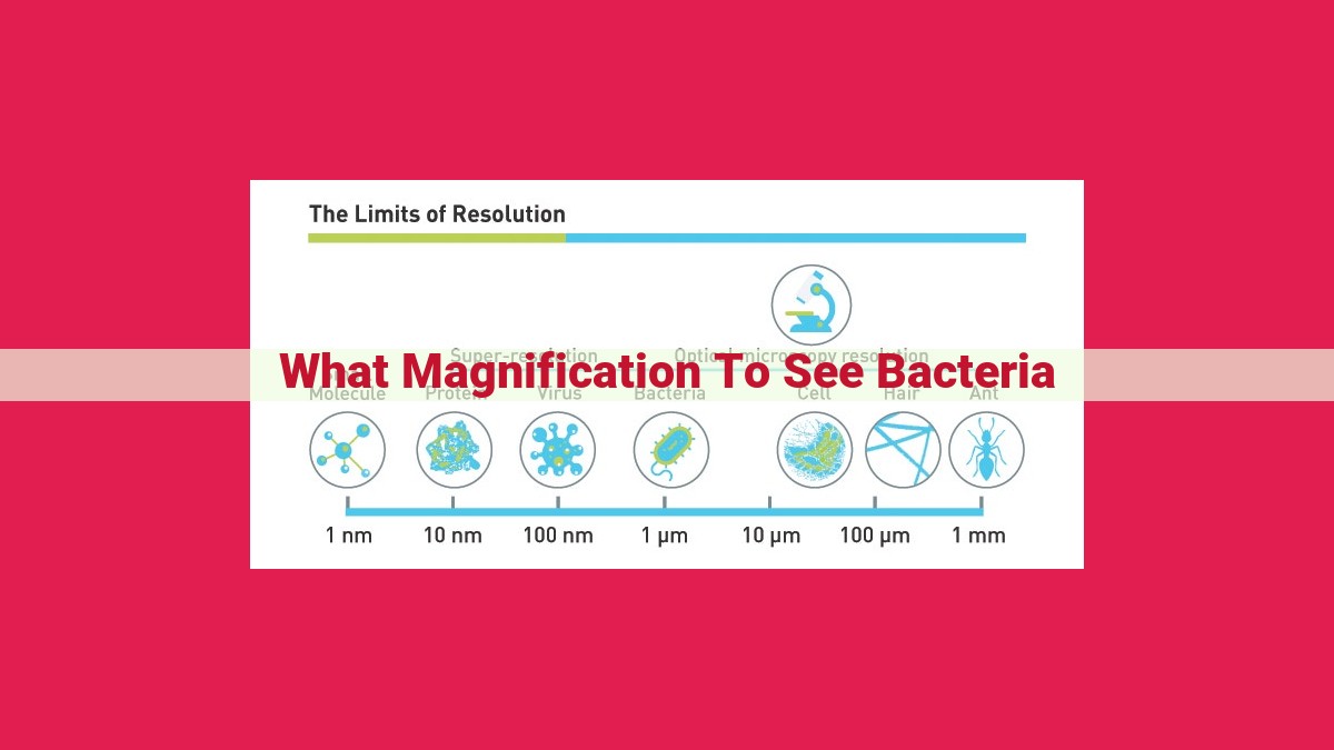
Understanding Bacterial Size and Resolution
Bacteria come in diverse shapes and sizes, ranging from tiny cocci to elongated bacilli. Their size determines the minimum magnification required to visualize them clearly. Resolution, on the other hand, refers to the ability to distinguish between two closely spaced objects. A higher resolution allows for greater detail to be observed.
Magnification Range for Optimal Observation
The ideal magnification range for bacterial observation typically falls between 400x and 1000x. This range provides a balance between image size and detail visibility. At lower magnifications, bacteria may appear too small for proper examination; at higher magnifications, resolution may be compromised, resulting in blurry and distorted images.
Consider the Desired Detail Level
The desired level of detail also influences the choice of magnification. For general morphological studies, a magnification of 400x may suffice. However, if specific structural features or cellular components need to be examined, higher magnifications such as 1000x become necessary.
Balancing Resolution and Magnification
It’s important to note that increasing magnification does not always result in better images. Resolution is a limiting factor in image clarity. If the objective lens does not have sufficient resolving power, increasing magnification beyond a certain point will only magnify the blur and reduce the overall image quality.
Choosing the Right Objective Lens
The objective lens is the key component that determines magnification and resolution. Each objective lens has a numerical aperture (NA), which indicates its light-gathering ability and, consequently, its resolving power. For optimal bacterial observation, objective lenses with NAs in the range of 0.65 to 1.4 are recommended.
By carefully considering bacterial size, resolution, and desired detail level, researchers can determine the optimal magnification for their observations. This empowers them to delve into the microscopic realm of bacteria, uncovering their hidden structures and shedding light on their fascinating world.
