Microscope Magnification, Resolution, And Field Of View: Understanding The Key Concepts

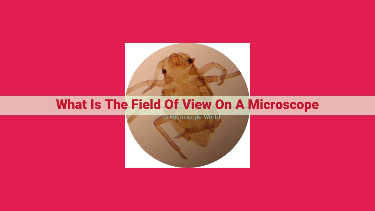
Field of view (FOV) is the circular area, visible through the microscope eyepiece, that contains the specimen. It is determined by the magnification of the eyepieces and objective lenses used. A larger FOV allows for a wider area to be observed, but reduces the overall magnification. Magnification refers to the ability of the microscope to enlarge the specimen, and is calculated by multiplying the magnification of the objective lens by that of the eyepiece lens. Resolution, on the other hand, is the ability to distinguish between two closely spaced objects. It is limited by the diffraction limit and the resolving power of the microscope.
Field of View: The Eye of the Microscope
In the realm of microscopy, field of view signifies the circular area visible through the microscope’s eyepiece. It plays a crucial role, determining the extent of the specimen you can observe at a glance. The eyepieces, typically with a magnification of 10x, and objective lenses, varying from 4x to 100x or more, work together to dictate the field of view.
Eyepieces and Objective Lenses: Shaping the View
The eyepiece, positioned at the top of the microscope, magnifies the image formed by the objective lens. Higher magnification eyepieces narrow the field of view, providing a more detailed view of a smaller area. Objective lenses, situated at the bottom of the microscope, gather light from the specimen and project an image onto the eyepiece. Lower magnification objective lenses produce a wider field of view, encompassing a larger portion of the specimen. As you switch to higher magnification objective lenses, the field of view shrinks, enabling you to focus on specific regions.
Understanding the interplay between eyepieces and objective lenses allows you to adjust the field of view to suit your observation needs. A wider field of view is ideal for scanning large specimens, while a narrower field of view is appropriate for examining minute details.
Magnification: Seeing the Unseen
In the realm of microscopy, magnification unveils the microscopic world, allowing us to explore the unseen depths of our surroundings. It transforms our perception, enabling us to delve into the intricate details of cells, organisms, and beyond.
Magnification, in essence, is the process of enlarging the apparent size of an object. When applied to microscopy, it empowers us to observe structures that are too tiny to be discerned by the naked eye. The key to magnification lies in the objective lenses of a microscope. These specialized lenses are designed to gather and focus light from the specimen, creating an enlarged image that we view through the eyepieces.
The total magnification of a microscope is determined by multiplying the magnification of the objective lens by the magnification of the eyepieces. For instance, if we use a 10x objective lens and 10x eyepieces, the resulting total magnification will be 100x. This means that the image we observe will be 100 times larger than its actual size.
Each objective lens bears its own specific magnification, ranging from low-power to high-power lenses. Low-power objectives provide a wider field of view, allowing us to scan larger areas of the specimen. High-power objectives, on the other hand, offer narrower fields of view but deliver greater magnification, enabling us to resolve finer details.
By adjusting the magnification, we can tailor our observations to match the desired level of detail. Low magnification facilitates a comprehensive examination, while high magnification grants us an in-depth exploration of specific structures. This versatility makes magnification a crucial aspect of microscopy, allowing us to probe the microscopic world with precision and efficiency.
Resolution: Unveiling the Finest Details
In the realm of microscopy, resolution emerges as a crucial parameter that defines the sharpness and clarity of the observed images. It represents the microscope’s ability to distinguish between finely spaced objects. Without sufficient resolution, intricate details may blur together, hindering accurate observation and analysis.
The fundamental principle behind resolution lies in the diffraction limit. This limit arises from the wave-like nature of light, which causes it to spread out as it passes through an aperture, such as the objective lens of a microscope. The smaller the aperture, the greater the diffraction, resulting in a lower resolution.
To quantify resolution, we introduce the concept of resolving power. Resolving power is defined as the ability to distinguish between two closely spaced objects. It is typically expressed as the minimum distance between two objects that can be seen as separate entities.
Factors Affecting Resolution:
-
Objective Lens Numerical Aperture (NA): The NA measures the light-gathering capacity of an objective lens. Lenses with higher NAs produce narrower cones of light, resulting in better resolution.
-
Wavelength of Light: Shorter wavelengths of light, such as blue light, have better resolving power than longer wavelengths, such as red light.
-
Immersion Oil: By using immersion oil between the objective lens and the specimen, the NA can be increased, leading to improved resolution.
Importance of Resolution:
High resolution is paramount in microscopy for a variety of reasons:
- Accurate identification and characterization of fine structures within cells and tissues
- Early detection of microscopic abnormalities
- Precise measurements and analysis of small biological specimens
- Imaging below the cellular level, enabling the visualization of viruses and macromolecules
Understanding the concepts of resolution, diffraction limit, and resolving power empowers researchers and practitioners to optimize their microscopy techniques, ensuring the most accurate and informative images.
Depth of Field: Focus on the Essentials
Unveiling the Microscopic Realm with Depth of Field
In the microscopic realm, capturing sharp and focused images is paramount. Depth of field plays a crucial role in achieving this precision. It refers to the range of distances within which objects appear sharp in an image. Understanding depth of field empowers microscopists to optimize their imaging techniques and reveal intricate details.
The Significance of Depth of Field
Depth of field is essential for microscopy as it allows researchers to:
- Distinguish objects at different elevations within the sample
- Visualize structures in three-dimensional perspective
- Obtain images with higher clarity and definition
Factors Influencing Depth of Field
Several factors influence the depth of field in microscopy:
Focal Plane
The focal plane is the plane within the sample that is in sharp focus. Depth of field is directly proportional to the thickness of the focal plane. A thicker focal plane results in a greater depth of field.
Working Distance
Working distance is the distance between the objective lens and the sample. Increasing the working distance decreases the depth of field. This is because a longer working distance results in a wider cone of light illuminating the sample, leading to a thinner focal plane.
Focus
The position of the focus within the sample also affects depth of field. Focusing on a plane closer to the objective lens reduces depth of field compared to focusing on a plane farther away. This is because a closer focus results in a smaller cone of light and a thinner focal plane.
Optimizing Depth of Field
To optimize depth of field for specific microscopy applications, researchers can adjust the following parameters:
- Choice of objective lens: Using a higher magnification objective lens (with a shorter working distance) decreases depth of field.
- Adjustment of working distance: Increasing the working distance slightly can increase depth of field, but excessive working distance can lead to reduced resolution.
- Fine-tuning focus: Precisely focusing on a specific plane within the sample can enhance depth of field.
Understanding depth of field is crucial for successful microscopy. By carefully considering the factors that influence depth of field, researchers can optimize their imaging techniques to obtain clear and insightful microscopic images. From revealing intricate cellular structures to capturing the dynamics of living organisms, depth of field empowers microscopists to explore the microscopic realm with unparalleled clarity.
Numerical Aperture: Unveiling the Unclear
In the realm of microscopy, numerical aperture (NA) stands as a crucial parameter that unveils the unseen. It is a quantitative measure of the light-gathering ability of an objective lens, and its significance lies in determining the resolution and depth of field of the microscope.
The condenser plays a pivotal role in shaping the numerical aperture. Its function is to concentrate light onto the specimen, increasing the illumination intensity and maximizing the amount of light that enters the objective lens. By manipulating the condenser’s position and aperture, researchers can optimize the NA for their specific imaging needs.
Another factor influencing NA is immersion oil. When applied between the objective lens and the specimen, immersion oil fills the air gap, reducing light scattering and effectively increasing the NA. This technique is particularly valuable for high-resolution imaging, as it enhances the ability to discern fine details.
The numerical aperture is directly proportional to the sinus of the half-angle of light acceptance by the objective lens. This means that lenses with larger NA values can collect more light and generate brighter images. Consequently, they offer improved resolution by allowing for tighter focusing and reduced diffraction effects.
Understanding the role of numerical aperture is essential for effective and efficient microscopy. By optimizing NA through proper condenser use and immersion oil techniques, researchers can maximize image quality, resolve finer structures, and delve deeper into the intricacies of their specimens.
Understanding Microscopy Concepts for Effective Exploration
In the realm of microscopy, a deep understanding of fundamental concepts is paramount for unlocking the mysteries of the unseen world. Mastering these principles empowers microscopists to optimize their observations, leading to accurate interpretations and groundbreaking discoveries.
Field of View: The Window to the Microcosm
The field of view is the circular area visible through the microscope’s eyepieces. It depends on the eyepieces and objective lenses used, each contributing to the total magnification. A wider field of view allows for a broader perspective, while a narrower field provides more detailed observation.
Magnification: Unveiling the Invisible
Magnification enlarges the apparent size of an object, making it easier to study. The objective lens is primarily responsible for magnification, with higher power objectives providing greater magnification. Total magnification is calculated by multiplying the magnifications of the objective and eyepiece lenses.
Resolution: Discerning Fine Details
Resolution determines the ability to distinguish between two closely spaced objects. It is limited by the diffraction limit, which arises from the wave-like nature of light. Higher numerical aperture objectives enhance resolution, allowing for finer details to be visualized.
Depth of Field: Focus on the Essential
Depth of field refers to the range of depths within a specimen that appear sharp. It depends on the focal plane, working distance, and focus adjustment. A large depth of field facilitates the examination of thick specimens, while a narrow depth of field isolates specific planes for detailed analysis.
Numerical Aperture: Illuminating the Unseen
Numerical aperture quantifies the light-gathering ability of an objective lens. It influences the brightness of the image and resolution. A higher numerical aperture allows for more light collection, resulting in brighter images and enhanced resolution.
These concepts form the cornerstone of effective microscopy. Understanding them enables microscopists to tailor their observations to specific research questions. By harnessing the power of field of view, magnification, resolution, depth of field, and numerical aperture, researchers can unlock the secrets of the microscopic world, advancing our knowledge and improving countless disciplines.