Lung Apices: Understanding Their Anatomy, Function, And Diagnostic Imaging

Lung apices are the superior portions of the lungs located above the clavicles, connecting to the thoracic cavity. They act as air cushions during breathing and protect underlying structures from impacts. These regions are vulnerable to diseases and can impact respiratory health. Chest X-rays and CT scans are used to visualize lung apices, aiding in diagnosis and monitoring.
Understanding Lung Apices: The Guardians of Our Breathing
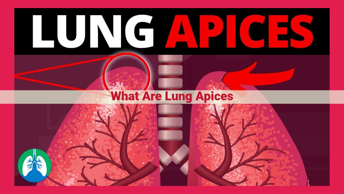
Lung apices, the dome-shaped tips of our lungs, play a crucial role in our respiratory system. Located just above our collarbones, these anatomical marvels connect to the thoracic cavity, the protective enclosure that houses our heart and lungs.
Anatomical Significance
Lung apices are composed of delicate lung tissue that extends upward into the superior thoracic aperture. They are surrounded by the pleura, a thin membrane that lines the lungs and thoracic cavity, creating a frictionless surface for lung movement. This unique positioning enables the apices to expand and contract during breathing.
Functional Wonders
Lung apices serve as air cushions during each inhalation, expanding to accommodate the increased volume of air entering our lungs. Moreover, they act as protective barriers, shielding the underlying structures in our thoracic cavity from external impacts.
Location and Structure: Unveiling the Secrets of the Lung Apices
Nestled atop the thoracic cavity, like two unsung heroes, reside the lung apices. These often-overlooked structures play a crucial role in the intricate symphony of respiration.
The lung apices are positioned gracefully above the clavicles, the prominent bones that form your collarbone. Like two upward-extending fingers, they reach into the thoracic cavity, the protective space that houses your lungs, heart, and other vital organs.
The lung apices are the uppermost extensions of the lungs, connecting seamlessly to the main bronchi, the primary airways that carry life-giving oxygen into the depths of your respiratory system. Each apex forms the very tip of a lung, providing a crucial point of contact between the respiratory organs and the surrounding structures.
As a result of their elevated positioning, the lung apices are surrounded by a unique set of anatomical neighbors. To the front, they border the superior mediastinum, a region that contains the heart and its major blood vessels. To the sides, they are embraced by the pleura, a thin membrane that lines the thoracic cavity and helps lubricate the movements of the lungs.
Functional Significance of Lung Apices
The lung apices, situated above the clavicles, play a crucial role in our respiratory system. They serve as a cushion of air during breathing, ensuring smooth lung expansion and contraction. This air-filled space acts as a buffer, reducing friction between the lungs and the surrounding structures.
Moreover, the lung apices offer protection to the delicate underlying structures, including blood vessels, nerves, and the thoracic cavity. In case of a sudden impact, the lung apices absorb a significant portion of the force, shielding these vital structures from damage. This protective role is particularly significant in safeguarding the heart, which lies directly beneath the apices.
Clinical Implications of Lung Apices: Susceptibility to Diseases and Impact on Respiratory Health
The lung apices, nestled snugly above our clavicles, play a crucial role in our respiratory system. However, their unique location makes them vulnerable to certain diseases and impacts respiratory health in complex ways.
Susceptibility to Tuberculosis and Lung Cancer
The lung apices are particularly susceptible to tuberculosis, a bacterial infection that primarily affects the lungs. This susceptibility stems from the thinness of the lung apices and their proximity to the cervical spine, which provides an easy pathway for bacteria to enter the thoracic cavity.
Similarly, lung cancer is more likely to develop in the lung apices due to their exposure to carcinogens present in tobacco smoke and air pollution. These toxins can accumulate in the lung apices over time, increasing the risk of malignant transformations within the lung tissue.
Impact on Respiratory Health
The location of the lung apices also influences respiratory health. During strenuous activities like running or exercising, the lung apices expand and compress to accommodate the increased airflow. This flexibility acts as an air cushion, protecting the underlying structures from sudden impacts and injuries.
However, lung apical disease can impair this protective function and lead to pain and discomfort during deep breathing. In severe cases, compromised lung apices can even impede oxygen intake and contribute to respiratory distress.
By understanding these clinical implications, healthcare professionals can better diagnose and treat lung apical diseases, safeguarding our respiratory well-being.
Diagnostic Evaluation of Lung Apices
Understanding the health of our lungs is crucial for overall well-being. The lung apices, located near the collarbone at the top of the thorax, play a pivotal role in respiration and protection. To assess the condition of these vital structures, medical professionals rely on advanced imaging techniques.
Chest X-rays provide a non-invasive method of visualizing the lung apices. This technique involves exposing the chest to a small amount of radiation, creating an image that can reveal abnormalities or structural changes. Chest X-rays aid in detecting conditions such as tuberculosis, lung cancer, and other respiratory diseases that may affect the apices.
Computed tomography (CT) scans offer a more detailed view of the lung apices. CT scans utilize multiple X-ray images from different angles to generate cross-sectional images. This technique allows physicians to examine the lung apices, surrounding structures, and potential abnormalities with greater precision. CT scans are particularly valuable in assessing complex conditions such as suspected tumors, fibrosis, and inflammatory processes that may impact the apices.
The findings obtained from chest X-rays and CT scans play a crucial role in diagnosing and monitoring lung diseases that affect the apices. Interpreting these images requires expertise, as subtle changes may indicate underlying health concerns. By analyzing the size, shape, and density of the lung apices, medical professionals can make informed assessments about their condition and potential treatments.
Regular diagnostic evaluations, including chest X-rays or CT scans, are often used to monitor the progression of lung diseases, assess _treatment response, and _identify potential complications. These imaging techniques provide invaluable insights into the health of the lung apices, ensuring timely diagnosis and appropriate management of respiratory conditions.