Interphase: Preparing The Cell For Mitosis (Comprehensive Guide)
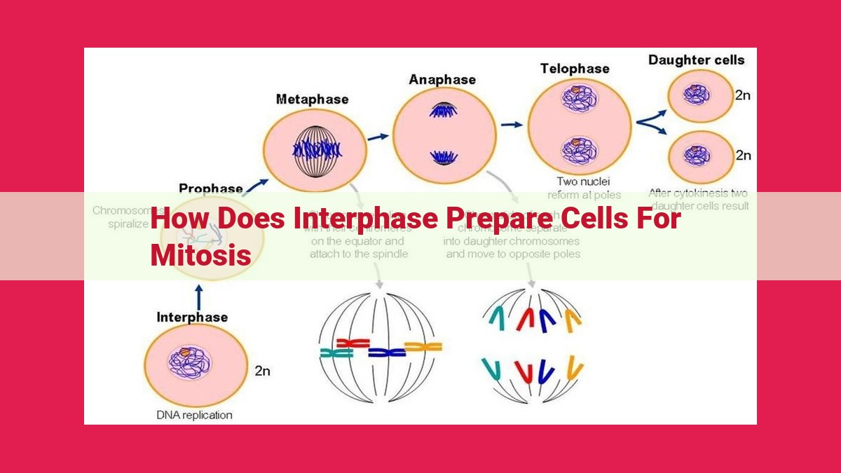
During interphase, the cell undergoes key processes preparing it for mitosis: DNA replication duplicates genetic material; centriole duplication ensures spindle fiber formation; nuclear envelope breakdown opens the command center, allowing spindle fibers to interact with chromosomes; and spindle fiber formation organizes chromosomes for orderly segregation during cell division.
DNA Replication: Copying the Genetic Blueprint for Life’s Journey
Imagine a world where every living organism carries a tiny instruction manual that guides its very existence. This manual is DNA, the molecule that holds the code for life. For cells to divide and create new life, this precious instruction book must be meticulously copied to ensure that each daughter cell receives an identical copy of the genetic blueprint.
Enter DNA replication, a remarkable process that unfolds during cell division. It is like a team of expert scribes working diligently to create an exact replica of the original text. First, the DNA molecule, shaped like a twisted ladder known as a double helix, unzips down the middle, exposing the individual strands. Then, enzymes called DNA polymerases step up to the plate, each one meticulously reading the sequence of bases (adenine, guanine, cytosine, and thymine) along one strand.
As the DNA polymerases move along the original strand, they match each base with its complementary partner. Adenine always pairs with thymine, and cytosine pairs with guanine. Like a master builder, they create a new strand of DNA that is complementary to the original template. As a result, each daughter cell that inherits a newly synthesized DNA molecule receives a complete and identical copy of the genetic blueprint. This process is critical for maintaining the integrity of the genetic code and ensuring the successful division of cells.
Centriole Duplication: The Unsung Heroes of Cell Division
As cells prepare for the momentous event of cell division, a crucial process takes place: centriole duplication. These tiny, cylindrical structures, located near the nucleus, play an indispensable role in organizing and orchestrating the cell’s dance of DNA segregation.
During interphase, the cell’s “resting” phase, centrioles replicate themselves in a highly controlled manner. Each mother centriole, consisting of nine triplets of microtubules, serves as a template for the assembly of a new daughter centriole. The daughter grows perpendicular to the mother, forming a right angle that will become the microtubule organizing center (MTOC) during cell division.
This duplication process ensures that each new cell, after division, inherits a complete set of centrioles. These structures are essential for the formation of spindle fibers, the “scaffolding” that aligns and separates the duplicated chromosomes during cell division. Without properly duplicated centrioles, the spindle fibers would not be organized correctly, leading to errors in chromosome segregation and potential genetic abnormalities.
Thus, the humble centrioles, often overlooked in the spotlight of DNA replication, play a vital role in the choreography of cell division. Their duplication during interphase lays the foundation for the precise and orderly distribution of genetic material to daughter cells, ensuring the continuity of life’s blueprints.
Nuclear Envelope Breakdown: Opening the Cell’s Command Center
The nuclear envelope, the membrane that encloses the cell’s nucleus, plays a crucial role in cell division. During mitosis, the nuclear envelope undergoes a significant transformation to prepare the cell for spindle fiber formation and chromosome separation.
In the early stages of mitosis, the nuclear envelope remains intact, protecting the cell’s genetic material from external interference. However, as the cell transitions to metaphase, the nuclear envelope begins to fragment and eventually dissolves. This breakdown is a precisely orchestrated event regulated by specific proteins that dismantle the envelope’s structure.
The breakdown of the nuclear envelope is essential for the formation of spindle fibers, which are microtubule structures that extend from the cell’s poles and align chromosomes for separation. With the nuclear envelope out of the way, spindle fibers can now penetrate the nuclear space and attach to the centromeres, the specialized regions of chromosomes that contain kinetochores. These attachments provide the physical link that allows spindle fibers to pull the chromosomes apart during anaphase.
The breakdown of the nuclear envelope also facilitates the activation of various cell cycle regulators that drive mitosis forward. Without the nuclear envelope acting as a barrier, these regulators can freely access their targets within the nucleus, ensuring that cell division proceeds smoothly and accurately.
Therefore, the breakdown of the nuclear envelope is a crucial step in mitosis, allowing spindle fibers to interact with chromosomes and initiating the events that lead to chromosome separation and cell division. This intricate process highlights the complexity and precision of cell biology, ensuring the faithful transmission of genetic material during cell reproduction.
Spindle Fiber Formation: Organizing the Chromosomes for Cell Division
As cells prepare to divide, they undergo a series of intricate processes to ensure that the genetic material is distributed equally to the daughter cells. One crucial step in this process is the formation of spindle fibers, which serve as the guiding tracks for chromosome movement during cell division.
Spindle fibers are composed of microtubule proteins that polymerize and depolymerize to form a dynamic network. These fibers extend from opposite poles of the dividing cell and attach to specific attachment points on the chromosomes called kinetochores.
The kinetochore is a protein complex that connects the spindle fibers to the chromosomes. It acts as the “checkpoint” of chromosome segregation, ensuring that each chromosome is properly attached to spindle fibers from both poles.
During metaphase, the chromosomes align at the center of the cell, with their kinetochores attached to spindle fibers from opposing poles. This alignment ensures that when the cell divides, each daughter cell will receive a complete set of identical chromosomes.
As the cell enters anaphase, the spindle fibers shorten, pulling the chromosomes apart. Each chromatid (identical copy of a chromosome) is separated and moved to opposite ends of the cell. This separation ensures that each daughter cell receives the same genetic content.
The formation of spindle fibers is a remarkable process that requires precise coordination and regulation. Disruptions in spindle fiber formation or function can lead to chromosome missegregation, which can result in genetic abnormalities and potential birth defects.
