The Importance Of Heat Fixation In Microscopy: Preservation And Accurate Analysis
Heat fixation is a crucial sample preparation technique in microscopy that involves exposing biological tissues to heat to enhance their preservation. It denatures and coagulates proteins, inactivating enzymes and preventing tissue autolysis. Thin tissue sections can then be obtained, minimizing artifacts and maintaining structural integrity. Counter staining adds contrast to different tissue components, aiding in their identification and interpretation. Heat fixation plays a vital role in preserving tissue integrity and providing a foundation for accurate microscopic analysis.
Heat Fixation: A Cornerstone of Biological Sample Preservation for Microscopy
In the realm of microscopy, preserving biological samples with utmost precision is paramount. Heat fixation, a fundamental technique, plays a crucial role in ensuring the integrity of these samples and unlocking their microscopic secrets.
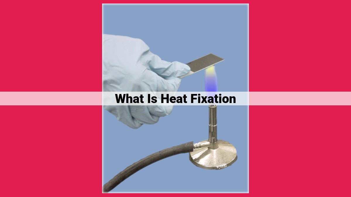
Heat fixation involves exposing biological specimens to elevated temperatures, typically 60-90 degrees Celsius. This process coagulates proteins, effectively immobilizing enzymes and preventing cellular autolysis, the destructive breakdown of tissues after death. By quenching destructive biochemical reactions, heat fixation preserves the cellular architecture, ensuring that the tissue remains intact and representative of its living state when examined under a microscope.
Heat Fixation: Preserving Specimens for Microscopic Examination
In the intricate realm of microscopy, heat fixation emerges as an indispensable technique that immortalizes biological specimens for meticulous observation. It plays a pivotal role in preserving the delicate integrity of tissues, capturing their intricate details, and revealing the secrets hidden within their structures.
Protein Transformations Under Heat’s Embrace
Heat fixation orchestrates a series of profound changes within the protein molecules that constitute cells and tissues. As temperatures rise, proteins undergo a process known as denaturation, where their carefully folded structures succumb to the disassembling forces of heat. This metamorphosis disrupts the intricate interactions between protein chains, leading to their coagulation into an insoluble, hardened mass.
Fixation: Halting Tissue’s Temporal Dance
The essence of heat fixation lies in its ability to arrest cellular processes, effectively pausing the relentless march of time within the specimen. By inactivating enzymes and preventing autolysis (the self-destruction of cells), heat fixation safeguards the integrity of tissue structures, preserving them in a state that accurately reflects their living counterparts.
This preservation extends beyond structural integrity, reaching into the realm of molecular dynamics. Heat fixation blocks enzymatic reactions and inhibits protein degradation, ensuring that the specimen’s biochemical composition remains intact. This meticulous conservation of both structure and molecular composition allows researchers to delve into the intricate workings of cells and tissues, unraveling their mysteries without the confounding effects of post-mortem changes.
Minimizing Artifacts: A Path to Clarity
Improper fixation techniques can introduce artifacts into tissue sections, distorting their appearance and potentially obscuring crucial details. Heat fixation, if executed skillfully, minimizes these imperfections, ensuring that the structures observed under the microscope accurately represent the true nature of the specimen.
Counter Staining: Enhancing the Visual Symphony
To further enhance the visibility and clarity of tissue components, counter staining is often employed in conjunction with heat fixation. This additional step introduces specific dyes that selectively bind to different cellular structures, creating a vibrant tapestry of colors that highlights specific features. This precise targeting enables researchers to differentiate between various cell types, identify subcellular structures, and unravel the intricate relationships that govern cellular organization.
Heat fixation stands as a cornerstone technique in the field of microscopy, providing researchers with a means to preserve and visualize biological specimens with remarkable fidelity. Its ability to arrest cellular processes, minimize artifacts, and facilitate counter staining empowers scientists to embark on groundbreaking discoveries, unraveling the mysteries of life at the cellular and subcellular levels.
Tissue Sections and Artifacts:
After heat fixation, tissue samples are meticulously sliced into thin sections to facilitate microscopic examination. These sections provide intricate glimpses into the cellular architecture of the sample. However, improper fixation techniques can lead to distortions or alterations, potentially compromising the accuracy of the microscopic analysis.
Distortions arise when heat fixation fails to adequately stabilize the tissue, leading to shrinkage, swelling, or the formation of voids within the tissue. These distortions can disrupt the normal arrangement of cells and make it challenging to draw accurate conclusions about the tissue’s structure and function.
Additionally, artifacts can occur as a result of suboptimal fixation. Artifacts are structures or changes that are not present in the actual tissue but appear as a consequence of the fixation process. Examples include coagulation artifacts, which arise from the excessive heat or prolonged fixation time, and extraction artifacts, where essential components of the tissue are dissolved and lost during the fixation procedure.
Understanding the potential pitfalls associated with heat fixation is paramount to ensure accurate and reliable microscopic observations. By carefully controlling the fixation process, pathologists can minimize distortions and artifacts, preserving the integrity of the tissue and maximizing the value of microscopic analysis for accurate diagnoses and research insights.
Counter Staining for Enhanced Microscopic Detail
In the realm of biological sample preparation, heat fixation plays a pivotal role in preserving tissue integrity. To further enhance the microscopic analysis of these samples, counter staining emerges as a valuable technique to reveal specific structures and improve visualization.
Counter staining involves the application of additional dyes to tissue sections, each targeting particular components or molecules. This process transforms the microscopic landscape, enabling the identification of diverse tissue elements with greater precision. By utilizing dyes with contrasting colors, structures of interest stand out against their surroundings, providing a clearer understanding of their distribution and morphology.
The benefits of counter staining extend beyond mere visual enhancement. It allows researchers to differentiate between various cell types, identify cellular components, and elucidate complex tissue architectures. Hematoxylin and eosin (H&E) stands as a classic example of counter staining, widely employed in histology to distinguish between the nucleus (stained blue) and cytoplasm (stained pink).
Counter staining techniques have revolutionized the field of microscopy, providing researchers with a powerful tool to explore the intricate details of biological samples. By harnessing the versatility of dyes and the precision they offer, counter staining empowers scientists to uncover hidden insights and gain a deeper understanding of the microscopic world.
