Human Somatic Cell Chromosomes: Structure, Mitosis, And Genetic Implications
Somatic cells, the body’s non-reproductive cells, contain 46 chromosomes in humans. This karyotype comprises 22 pairs of autosomes (non-sex chromosomes) and one pair of sex chromosomes (XX in females, XY in males). During mitosis, the process by which somatic cells divide, chromosomes condense and become visible as distinct “X” shapes in prophase. Each chromosome consists of two chromatids joined at the centromere, and its DNA is packaged into nucleosomes. Heterochromatin and euchromatin regions along the chromosome reflect differences in gene activity, while telomeres protect chromosome ends.
The Fascinating World of Human Chromosomes: Unraveling the 46-Chromosome Mystery
The Karyotype: A Blueprint of Chromosomal Identity
Imagine a jigsaw puzzle, each piece representing a chromosome. Your karyotype is a complete picture of this chromosomal puzzle, revealing the number, size, and shape of your chromosomes. It’s like a DNA fingerprint, providing crucial information about your genetic makeup.
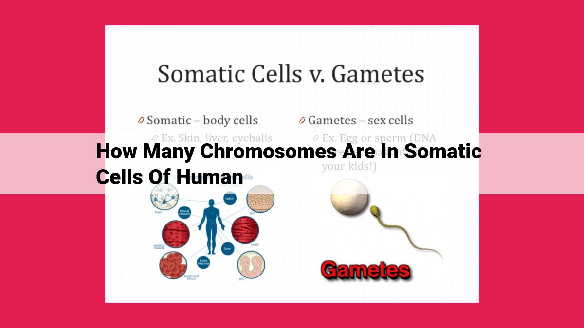
Autosomes vs. Sex Chromosomes: The Building Blocks of Life
Your karyotype consists of autosomes and sex chromosomes. Autosomes are the non-gender-specific chromosomes, present in both males and females. We inherit 23 pairs of autosomes, one set from our mother and one from our father. Sex chromosomes, on the other hand, determine biological sex. Females have two X chromosomes (XX), while males have an X and a Y chromosome (XY).
The 46-Chromosome Enigma: A Perfect Balance
The total number of chromosomes in human somatic cells (non-reproductive cells) is 46. This precise count is maintained through cell division: During mitosis, each daughter cell receives an identical set of chromosomes, ensuring the stability of genetic information across generations.
Cell Division and Somatic Cells: Understanding the Blueprint of Life
Every living organism, from the tiniest bacteria to the colossal blue whale, owes its existence to the intricate symphony of cells. Within each cell resides a microscopic library of genetic information encoded in the form of chromosomes. Somatic cells, the building blocks of our bodies, possess a distinct number of chromosomes that play a crucial role in cell division and the development of the organism.
The Miracle of Mitosis
The life cycle of somatic cells is governed by the process of cell division, known as mitosis. This remarkable dance of genetic material ensures that each new cell receives an identical copy of the parent cell’s chromosomes. Mitosis consists of several stages, each with a specific function:
1. Interphase: The cell grows and prepares for division by replicating its chromosomes.
2. Prophase: The replicated chromosomes condense and become visible, forming the distinctive “X” shape. The nuclear envelope breaks down, revealing the chromosomes.
3. Anaphase: The mitotic spindles, made of microtubules, attach to the centromere of each chromosome and pull the sister chromatids apart, moving them to opposite poles of the cell.
4. Telophase: The chromosomes reach the poles and start to unwind. Two new nuclear envelopes form around the chromosomes, and the cell membrane pinches in the middle, dividing into two daughter cells.
The Significance of Chromosomes in Somatic Cells
The precise number of chromosomes in somatic cells is essential for maintaining the organism’s genetic integrity. In humans, for instance, somatic cells contain 46 chromosomes, organized into 23 pairs. These pairs consist of one chromosome inherited from each parent. The importance of this karyotype, or chromosomal arrangement, extends to the proper development and functioning of the organism throughout its life.
Chromosome Visibility during Cell Cycle
- Introduction of centromere and chromatids
- Explanation of when chromosomes become visible (prophase) and the distinctive “X” shape they form
Chromosome Visibility during the Cell Cycle: A Glimpse into Cellular Dynamics
Within the intricate realm of cells, chromosomes, the carriers of our genetic heritage, play a pivotal role. While often shrouded in the invisible depths of the nucleus, these vital structures reveal their presence during specific stages of the cell cycle.
Centromere and Chromatids: The Structural Framework
Each chromosome consists of a centralized structure known as the centromere. It acts as the anchoring point for the two identical copies of DNA called chromatids. Imagine the centromere as a tiny bridge connecting two arms, each arm being a chromatid.
Prophase: The Unveiling Act
As the cell prepares for division, it embarks on a journey called prophase. During this phase, chromosomes begin to condense and shorten, becoming visible under a microscope. It’s like the blueprints of your life emerging from the shadows.
X-Shaped Formation: A Visual Cue
As the chromatids become even more visible, they align side by side, forming the distinctive “X” shape that is characteristic of chromosomes during prophase. This X-shaped appearance reflects the fact that each chromosome consists of two identical copies of DNA.
Unraveling the Mystery of Chromosome Visibility
The visibility of chromosomes during prophase is crucial for the cell’s ability to ensure accurate distribution of genetic material during cell division. The X-shaped formation and the centromere play a vital role in aligning the chromosomes and ensuring their proper segregation.
Exploring Other Aspects of the Cell Cycle
While this article focuses on chromosome visibility during the cell cycle, other stages also hold fascinating insights. Join us in future explorations as we delve into the intricacies of mitosis, meiosis, and the dynamic world of cell division.
Chromosome Structure: Unraveling the Building Blocks of Life
Every cell in our bodies houses a microscopic world of intricate machinery, and at the heart of this cellular landscape lie our chromosomes – the blueprints that carry the secrets of our genetic inheritance. The structure of these chromosomes tells a tale of meticulous organization and sophistication that has fascinated scientists for centuries.
Nucleosomes: The Master Organizers of DNA
Imagine a vast library filled with books that hold immense knowledge. The challenge lies in keeping these books organized and accessible. In the world of chromosomes, nucleosomes play a similar role. These protein structures wrap around DNA like tiny spools, forming a beaded chain that compacts the genetic material into a manageable form.
Telomeres: The Guardians of Chromosome Integrity
At the ends of each chromosome are specialized caps called telomeres. These regions of repetitive DNA are likened to the plastic tips of shoelaces, preventing the ends of the chromosomes from unraveling and fusing with neighboring ones. As essential as telomeres are to maintaining chromosome stability, they also mark the passage of time. With each cell division, telomeres gradually shorten, reflecting the limited lifespan of our cells.
Heterochromatin and Euchromatin: Regions of Secrets and Activity
Chromosomes are not uniform structures. They feature regions known as heterochromatin and euchromatin, each with its distinct characteristics. Heterochromatin, condensed and tightly packed, contains DNA that remains mostly silent and inactive during gene expression. In contrast, euchromatin is a more open and accessible region where genes are actively transcribed into RNA, providing the instructions for protein synthesis and cellular function.
By understanding the intricate structure of chromosomes, we gain deeper insights into the mechanisms that govern cell division, ensure the faithful transmission of genetic information, and protect the integrity of our DNA from damage. These insights empower us to unravel the mysteries of life and open up new avenues for diagnosing and treating diseases.
