Optimized Seo Title:unlock The Power Of The Gram Stain: Unveiling Bacterial Secrets And Empowering Diagnostics

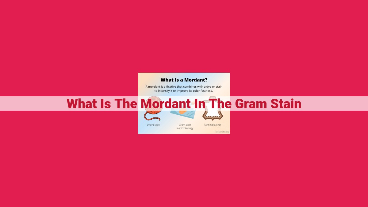
In the Gram stain, the mordant (often iodine) plays a crucial role as a dye-target material binder. By interacting with crystal violet, the primary stain, the mordant enhances its binding to the cell wall. This complex formation is then stabilized by Gram’s iodine, preventing its removal during decolorization. The mordant thus aids in distinguishing Gram-positive bacteria, which retain the crystal violet-iodine complex, from Gram-negative bacteria, which lose it due to their outer membrane structure. The Gram stain is a valuable technique for bacterial classification, aiding in diagnostic procedures and guiding antibiotic selection.
The Curious Case of the Mordant in the Gram Stain
In the fascinating world of microbiology, one technique reigns supreme for differentiating bacteria: the Gram stain. At the heart of this discovery lies a remarkable chemical called the mordant, a true maestro in the art of staining.
Imagine a dye, like crystal violet, eager to adhere to bacterial cell walls. However, some bacteria, the Gram-negative kind, have a protective outer membrane that keeps the dye at bay. Enter the mordant, a clever agent that acts as a bridge between the dye and the cell wall, allowing crystal violet to penetrate and paint the bacterium a deep purple.
The mordant, typically a metal salt like iodine, achieves this by forming a complex with the dye, creating an irresistible duo. This complex then latches onto the cell wall, strengthening the bond between the dye and the bacterium.
Now, let’s explore how the mordant contributes to the Gram stain’s success:
Purpose of the Gram Stain: Unraveling Bacterial Secrets
The Gram stain is a crucial technique for classifying bacteria into two distinct groups: Gram-positive and Gram-negative. This distinction is based on the bacteria’s response to the stain, which reveals differences in their cell wall structure.
Crystal Violet: The Primary Stain
Crystal violet, the primary stain in the Gram stain, is drawn to the mordant-treated cell walls of both Gram-positive and Gram-negative bacteria. However, when alcohol, the decolorizing agent, is introduced, it weakens the mordant-crystal violet complex in Gram-negative bacteria, allowing the dye to escape and leaving them unstained.
Mordant’s Role: Enhancing Crystal Violet Binding
The mordant plays a pivotal role in ensuring that the crystal violet binds effectively to Gram-positive bacteria. Its presence strengthens the interaction between the dye and the cell wall, preventing the dye from being washed away during the decolorization step. As a result, Gram-positive bacteria retain their purple color, a sign of their thick, peptidoglycan-rich cell walls.
The mordant is an unsung hero in the Gram stain, enhancing crystal violet binding and enabling the differentiation of Gram-positive and Gram-negative bacteria. Its role in this diagnostic technique has revolutionized the field of microbiology, providing a powerful tool for understanding and classifying bacteria.
Gram Stain: Delving into the Art of Bacterial Classification
In the realm of bacteriology, the Gram stain stands as a cornerstone technique for distinguishing between two distinct groups of bacteria: Gram-positive and Gram-negative. This staining method not only aids in their identification but also provides valuable insights into their susceptibility to antibiotics and other treatments.
The Gram stain owes its effectiveness to a crucial component known as the mordant. This substance serves as a dye-target material binder, essentially preparing the bacterial cell wall for enhanced crystal violet binding. Crystal violet, the primary stain in the Gram stain, imparts a deep purple hue to the bacteria and plays a significant role in differentiating between the two groups. Gram-positive bacteria retain this purple color, while Gram-negative bacteria lose it during the subsequent steps of the staining procedure.
The mordant’s role in this process becomes evident when we delve into the structural differences between Gram-positive and Gram-negative bacteria. Gram-positive bacteria possess a thick peptidoglycan layer in their cell walls, which enables them to bind more readily to the crystal violet-mordant complex. In contrast, Gram-negative bacteria have a thinner peptidoglycan layer and an additional outer membrane that acts as a permeability barrier, preventing the retention of crystal violet.
During the Gram stain procedure, the mordant facilitates the formation of a crystal violet-iodine complex within the cell wall, solidifying the bond between the dye and the bacterial surface. This complex is then stabilized by Gram’s iodine, which acts as a fixative. The subsequent application of alcohol, a decolorizing agent, dissolves the outer membrane of Gram-negative bacteria, allowing the crystal violet-iodine complex to escape. This step effectively decolorizes Gram-negative bacteria, while Gram-positive bacteria remain purple.
To complete the process, a counterstain, such as safranin, is employed to stain both Gram-positive and Gram-negative bacteria a contrasting color, typically red. This aids in visualizing all bacteria under the microscope, providing a comprehensive view of the microbial sample.
Understanding the role of the mordant and the sequence of steps involved in the Gram stain is crucial for its accurate interpretation. This technique not only serves as a diagnostic tool for bacterial classification but also guides appropriate antibiotic selection and contributes to our understanding of bacterial pathogenesis.
Crystal Violet: The Primary Stain in the Gram Stain Technique
As we embark on the fascinating journey of bacterial classification, we encounter a technique that has revolutionized our understanding of these microscopic organisms: the Gram stain. Crystal violet, the primary stain, plays a pivotal role in this technique, revealing the secrets of bacterial structure and behavior.
Crystal violet is a triphenylmethane dye, characterized by its violet color. It possesses a unique affinity for cell walls that have been pretreated with a mordant, a substance that enhances the dye’s adherence. This mordant, typically iodine, reacts with the peptidoglycan layer of Gram-positive bacterial cell walls, providing a binding site for the crystal violet.
Once the cell wall is receptive, the crystal violet seeps into its porous structure, impregnating the Gram-positive bacterial cell wall and firmly attaching to the peptidoglycan layer. This binding results in the retention of the violet stain, even after subsequent decolorization steps. In contrast, the outer membrane of Gram-negative bacteria prevents the crystal violet from accessing the peptidoglycan layer, leading to its removal during decolorization.
Thus, the crystal violet stain serves as a cornerstone of the Gram stain technique, differentiating Gram-positive from Gram-negative bacteria based on their cell wall structures. This distinction is crucial in bacterial identification, diagnosis, and treatment, guiding appropriate antibiotic selection and infection management strategies.
Gram’s Iodine: The Keeper of the Purple Crystal
In the fascinating world of microbiology, the Gram stain stands as a powerful technique for distinguishing between two major bacterial groups: Gram-positive and Gram-negative. At the heart of this distinction lies a crucial component known as Gram’s iodine, an unsung hero that plays a pivotal role in securing the purple crystal, crystal violet, to the bacterial cell wall.
The Power of Gram’s Iodine: A Fixative for the Ages
Imagine the crystal violet-iodine complex within the bacterial cell wall as an exquisite tapestry. Gram’s iodine acts like a master weaver, its function to bind the intricate threads of this complex together, creating an indissoluble bond that resists the onslaught of time. Once established, this complex becomes a testament to the artistry of Gram’s iodine, unyielding to the forces that seek to disrupt its existence.
Preventing the Great Dye Escape: The Significance of Fixation
The strength of the crystal violet-iodine complex is paramount, for it ensures the successful differentiation of Gram-positive and Gram-negative bacteria. Without the firm grip of Gram’s iodine, the dye complex would fall apart prematurely during the decolorization step, rendering the Gram stain less effective. In this sense, Gram’s iodine serves as the backbone of the technique, the unsung hero that guarantees the validity of our bacterial classification.
A Diagnostic Tool of Unrivaled Importance
The Gram stain, with Gram’s iodine as its steadfast companion, has revolutionized the field of microbiology. It provides a rapid, reliable method for categorizing bacteria, enabling clinicians to make informed decisions regarding treatment and infection control. The diagnostic value of the Gram stain cannot be overstated, and it owes its success in no small part to the unwavering presence of Gram’s iodine.
Like an invisible thread, Gram’s iodine weaves its magic within the Gram stain, solidifying the crystal violet-iodine complex and preventing its untimely demise. Its role, though understated, is indispensable, ensuring the accuracy and reliability of this cornerstone technique. As we continue to explore the microbial world, let us remember the silent yet vital contribution of Gram’s iodine, the guardian of the purple crystal.
Alcohol: Decolorizing Agent
- Purpose in removing the crystal violet-iodine complex from Gram-negative bacteria.
- Mechanism of dissolving the outer membrane and allowing dye escape.
Alcohol: The Dye Remover in the Gram Stain
In the Gram staining technique, alcohol plays a pivotal role as the decolorizing agent, responsible for differentiating between Gram-positive and Gram-negative bacteria. This crucial step in the process selectively removes the crystal violet-iodine complex from the cell walls of Gram-negative bacteria, allowing us to distinguish them from their Gram-positive counterparts.
The alcohol used in the Gram stain is typically a mixture of ethanol and acetone. It functions by dissolving the lipid-rich outer membrane of Gram-negative bacteria, which acts as a barrier to the crystal violet-iodine complex. This dissolution creates a permeable pathway through which the dye complex can escape from the cell wall, causing the bacteria to be decolorized.
In contrast, Gram-positive bacteria have a thick peptidoglycan layer in their cell walls, which lacks the lipid-rich outer membrane found in Gram-negative bacteria. This difference in cell wall structure prevents the alcohol from dissolving the peptidoglycan layer, and the crystal violet-iodine complex remains trapped within the cell wall, resulting in the bacteria remaining purple.
Therefore, alcohol is an indispensable component of the Gram stain technique. Its ability to selectively remove the crystal violet-iodine complex from Gram-negative bacteria distinguishes them from Gram-positive bacteria, aiding in the identification and classification of these microorganisms. This differentiation is essential in the diagnosis and treatment of various bacterial infections, making the Gram stain a vital tool in medical microbiology.
Safranin: Counterstain
- Properties of the red-colored dye and its ability to stain both Gram-positive and Gram-negative bacteria.
- Staining mechanism by impregnating the cytoplasm and highlighting all bacteria.
Safranin: The Color that Unifies
As we conclude our exploration of the Gram stain technique, let’s delve into the final component: safranin, the counterstain that brings color to our bacterial tapestry. This red-hued dye possesses a remarkable ability to stain both Gram-positive and Gram-negative bacteria.
Unlike crystal violet, safranin doesn’t discriminate. Its non-specific binding mechanism allows it to penetrate the cytoplasm of all bacteria, regardless of their Gram reaction. Once inside, safranin imparts a uniform red color, highlighting the presence of all bacteria in the sample.
This contrasting color serves as a visual cue, complementing the distinction made by crystal violet. While Gram-positive bacteria remain purple, Gram-negative bacteria, after being decolorized, take on the red hue of safranin. It’s like two contrasting shades painting a vivid picture of microbial diversity.
In the diagnostic world, the Gram stain is a crucial tool for classifying bacteria. By employing a series of specific reagents, including safranin, microbiologists can gain valuable insights into the nature of bacterial infections. This information guides appropriate treatment plans and helps ensure the well-being of patients.