Understanding The Eye: How Light And Structures Work Together For Vision

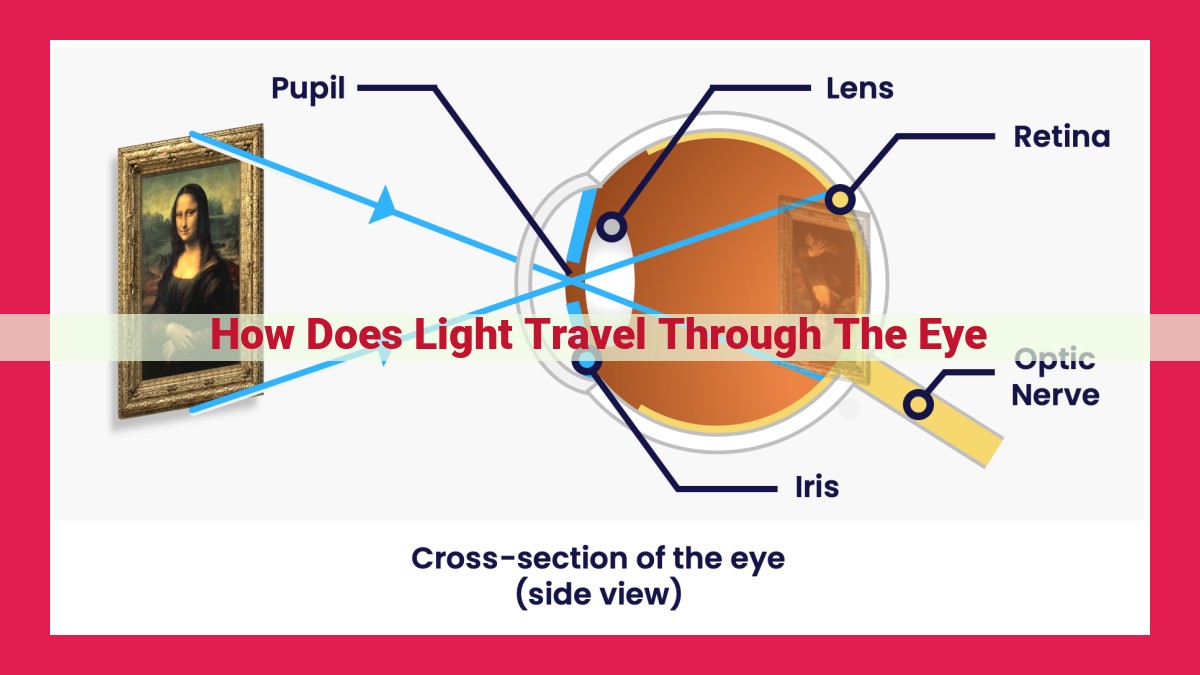
Light enters the eye through the cornea and pupil, refracting to focus on the retina. The retina contains photoreceptors that convert light into electrical signals, which travel through the optic nerve to the brain. The lens adjusts its shape to fine-tune focus, while the vitreous humor supports the eye’s structure. The iris controls pupil size to regulate light intake.
The Journey of Light: Unraveling the Eye’s Anatomy
Our eyes are portals to a vibrant world, transforming light into the images that shape our reality. Understanding the intricate anatomy of the eye is key to appreciating the marvel of this process and safeguarding our precious vision.
The Protective Shield: The Cornea
The cornea, the transparent outermost layer of the eye, resembles a crystal shield. It refracts light, ensuring crisp focus on the retina, while also acting as a protective barrier against external elements. Its smooth, dome-like surface allows light to enter the eye without distortion.
The Gateway to the Eye: The Pupil
Imagine the pupil as a portal, the black opening in the center of the iris. This dynamic structure adjusts its size to regulate the amount of light entering the eye. In bright environments, the pupil constricts, while in dim conditions, it widens, like the iris of a camera.
The Colorful Controller: The Iris
The iris, the colored part of the eye, adds beauty to the face but also plays a vital role. It surrounds the pupil and controls its diameter, influencing both eye color and the amount of light entering the eye. Pigments within the iris give it its unique hue, creating the diversity of eye colors we observe.
The Precision Tool: The Lens
Behind the iris lies the lens, a flexible, crystalline structure that further refines the incoming light. By changing its shape, the lens ensures that light is focused precisely on the retina. This remarkable ability allows us to clearly view objects at varying distances.
The Cushioning Support: The Vitreous Humor
Filling the main cavity of the eye, the vitreous humor acts as a cushioning gel, supporting the lens and maintaining the eye’s shape. This transparent substance provides a stable environment for the delicate inner structures, preserving the eye’s delicate balance.
The Sensory Canvas: The Retina
The retina, a thin, light-sensitive layer at the back of the eye, is where the magic of vision unfolds. It contains photoreceptor cells specialized in detecting light and converting it into electrical signals that the brain can interpret as images.
The Light Detectors: Photoreceptors
Within the retina, photoreceptors function as the eye’s tiny light detectors. These specialized cells respond to different wavelengths of light, allowing us to perceive the full spectrum of colors. Rods, sensitive to low light, aid in night vision, while cones facilitate color vision and detail.
The Visual Connector: The Optic Nerve
The optic nerve, a bundle of nerve fibers, connects the retina to the brain’s visual cortex. It transmits the electrical signals generated by the photoreceptors, carrying visual information to the brain, where it is processed into the images we see.
**The Cornea: The Eye’s Protective Shield**
Nestled at the forefront of your visual journey, the cornea emerges as a translucent, dome-shaped shield guarding the delicate inner workings of your eye. This remarkable structure, the cornea, is your eye’s first defense against the world, providing a clear path for light to reach your retina and transform into a symphony of colors and shapes.
As the light embarks on its odyssey into your eye, it encounters the cornea as its first challenge. With the precision of a skilled lensmaker, the cornea deftly refracts and focuses the incoming light, bending its path to ensure a sharp image on your retina, the sensory canvas within.
Beyond its role as a light-bending marvel, the cornea stands as a staunch protector of your eye’s delicate interior. Its tightly packed collagen fibers form a nearly impenetrable barrier, shielding the inner eye from dust, debris, and harmful microorganisms that could disrupt the delicate visual symphony within.
Keywords: #cornea, #eye protection, #light refraction, #visual acuity, #eye anatomy
The Pupil: The Gateway to the Eye
In the intricate tapestry of the eye, the pupil stands as a resilient sentinel, safeguarding your vision’s journey. This enigmatic black opening, nestled amidst the iris’s vibrant hues, serves as a gateway for light to penetrate the depths of this remarkable organ and unveil the world around you.
Like an ever-vigilant guardian, the pupil adapts seamlessly to the ebb and flow of light conditions. In bright environments, it contracts, reducing the amount of illumination entering the eye to prevent overexposure and protect the sensitive retina within. Conversely, as darkness descends, the pupil expands, allowing more light to enter and enhancing your ability to navigate dimly lit spaces.
This remarkable adaptation is orchestrated by the muscles within the iris, known as the sphincter and dilator pupillae. When the sphincter pupillae contracts, the pupil narrows, reducing the aperture through which light can enter. Conversely, the dilator pupillae relaxes, resulting in pupil dilation.
The pupil’s size also plays a crucial role in controlling the depth of field. By altering the dimensions of this aperture, the eye can fine-tune its focus on objects at varying distances. When the pupil is small, it provides greater depth of field, allowing you to see objects both near and far with equal clarity. In contrast, a large pupil reduces depth of field, enhancing your focus on objects closer to the eye while blurring those in the background.
So, as your eyes behold the world, remember the unassuming yet pivotal role of the pupil. It is the gateway through which light transforms into a symphony of colors, shapes, and memories, illuminating your visual experience and connecting you to the wonders of sight.
The Iris: The Colorful Controller of the Eye
Imagine your eye as a sophisticated camera, and the iris is its intricate lens—the iris, the vibrant part of your eye that surrounds the pupil, plays a crucial role in controlling light and giving you your unique eye color.
The iris is an exquisite masterpiece, not only for its captivating hues but also for its crucial function in regulating the amount of light entering your eye. Think of it as an adjustable diaphragm in your camera. Thanks to tiny muscles in the iris, it can rapidly expand or contract, adjusting the size of the pupil, the black circular opening in the center. By controlling the pupil’s diameter, the iris decides how much light reaches the retina, the light-sensitive layer at the back of your eye, which translates light into electrical signals that your brain interprets as images.
So, how does the iris influence your eye color? It’s all thanks to melanin, the same pigment responsible for your skin and hair color. Melanin resides within the iris, and its concentration determines your eye’s unique shade. Melanin-rich irises appear brown or black, while those with less melanin appear blue or green.
The remarkable ability of the iris to control the size of the pupil is essential for clear vision. In bright sunlight, for instance, the iris automatically constricts the pupil to reduce the amount of light entering the eye, protecting your retina from damage. Conversely, in dim environments, the iris dilates the pupil, allowing more light to reach the retina and enhance your vision in low-light conditions.
In essence, the iris is an intricate masterpiece, both visually and functionally. It’s a testament to the sophistication of the human body, regulating light, protecting the retina, and adding a touch of vibrant color to our vision. So, the next time you look into someone’s eyes, take a moment to appreciate the iris, the colorful controller that orchestrates the symphony of vision.
The Lens: The Precision Tool of Vision
Behind the pupil and iris lies the lens, a remarkable structure that plays a pivotal role in the eye’s intricate symphony of light. This flexible, transparent organ is responsible for fine-tuning the focus of incoming light, ensuring that images are sharp and clear on the retina.
The lens is a dynamic structure that is capable of changing its shape, much like a skilled artisan molding clay. This ability to alter its curvature allows the lens to focus light onto the retina with remarkable precision. The process is known as accommodation, and it’s essential for clear vision at different distances.
Imagine holding a magnifying glass close to an object. As you move the glass closer, the image becomes larger. Similarly, when the eye focuses on a nearby object, the lens assumes a more rounded shape, increasing its refractive power. This allows the incoming rays of light to bend more sharply, bringing the image into focus on the retina.
Conversely, when the eye gazes at a distant object, the lens flattens out, decreasing its curvature. This reduces its refractive power, allowing the incoming light rays to converge more gradually, again landing the image sharply on the retina.
The lens’s ability to adapt is made possible by tiny muscle fibers called the ciliary muscles. These muscles surround the lens and contract or relax under the control of the nervous system. By adjusting the tension on the lens, the ciliary muscles alter its shape, enabling the eye to focus smoothly from near to far and back again.
The lens is truly a marvel of biological engineering. Its ability to change shape with such precision is essential for clear vision and our ability to navigate the world around us.
The Vitreous Humor: The Eye’s Cushioning Support
Nestled in the cavernous depths of your eye, lies a remarkable gelatinous substance known as the vitreous humor. A transparent, gel-like enigma, it plays a pivotal role in maintaining the shape and health of your precious vision.
Maintaining Structural Integrity
Like a resilient cushion, the vitreous humor fills the vitreous chamber, the main cavity of your eye, accounting for about two-thirds of its volume. Its viscous nature contributes to the eye’s overall shape, ensuring it maintains its spherical form. This stable structure provides an ideal environment for the proper functioning of the delicate tissues within.
Supporting the Lens
The vitreous humor also serves as a supportive cradle for the lens, the delicate structure responsible for focusing light on the retina. As the lens changes shape to adjust focus, the vitreous humor gently cushions and suspends it, ensuring its precise positioning.
Maintaining the Eye’s Transparency
Light effortlessly traverses the transparent vitreous humor, allowing it to reach the retina without distortion. This crystalline clarity is crucial for clear and sharp vision.
Protecting Inner Structures
In addition to providing structural support, the vitreous humor also acts as a protective barrier. Its gelatinous consistency prevents the delicate retina from coming into contact with the other components of the eye, shielding it from potential damage.
Ongoing Renewal
The vitreous humor, like all living tissues, is constantly renewed. Fresh vitreous humor is produced by the ciliary body, a ring-shaped structure located behind the iris. As new vitreous humor forms, it gradually replaces the older vitreous humor, ensuring the eye remains healthy and functional throughout life.
The Retina: The Sensory Canvas
Nestled at the back of your eye, the retina is an intricate tapestry of cells, the foundation of your vision. Imagine it as nature’s camera film, capturing every scene, every detail, and painting it into the images we perceive.
Within this thin, light-sensitive layer, millions of photoreceptor cells await the kiss of light. These specialized cells, like tiny sentinels, stand ready to convert the photons of light into electrical impulses. It’s a remarkable symphony where the language of light is translated into the language of the brain.
Cones and rods, two distinct types of photoreceptors, share the canvas. Cones, sensitive to color and detail, excel in bright light, revealing the vibrant world we see in daylight. Rods, on the other hand, reign supreme in dim conditions, guiding us through the mysteries of night.
They respond to the slightest flickers of light, triggering a chain reaction that transforms visual information into electrical signals. These signals then embark on a journey through the optic nerve, a bundle of fibers that carries them to the brain. There, in the visual cortex, the electrical impulses are interpreted, painting the scenes we witness with eyes wide open.
Photoreceptors: The Light Detectors
Within the intricate tapestry of the eye, there lie specialized cells known as photoreceptors, the gatekeepers of our visual experience. These remarkable cells possess an astonishing ability to transform the symphony of light that reaches our eyes into a pulsating language of electrical impulses, which are then transmitted to the brain, where they are deciphered and interpreted as the world we see.
Photoreceptors are meticulously arranged within the retina, the light-sensitive lining at the back of the eye. These tiny cells are divided into two main types: cones and rods. Cones allow us to perceive colors and fine details in well-lit environments, while rods excel in low-light conditions, enabling us to navigate the shadows and perceive shapes and movements.
The extraordinary sensitivity of these cells is a testament to their ingenious design. Each photoreceptor contains a pigment molecule called retinal, which is nestled within a membrane. When light strikes the retinal molecule, it undergoes a subtle transformation, triggering a cascade of molecular events that ultimately generate an electrical impulse.
The electrical signals generated by the photoreceptors are then relayed to the optic nerve, a bundle of nerve fibers that connects the retina to the brain. These signals travel along the optic nerve to the visual cortex, a specialized region of the brain responsible for interpreting visual information.
The intricate interplay between photoreceptors, the optic nerve, and the visual cortex forms the foundation of our vision. Without these remarkable cells, we would be unable to appreciate the vibrant colors of a sunset, navigate the intricacies of our surroundings, or experience the wonders of the visual world.
The Optic Nerve: The Visual Connector
The optic nerve serves as the crucial bridge between the eye’s sensory realm and the brain’s interpretative center. Imagine it as a tireless courier, carrying vital messages from the retina to the brain’s visual cortex.
Imagine light entering the eye, painting a vibrant canvas on the retina. These images are captured by specialized photoreceptor cells, which convert them into electrical impulses. The optic nerve then steps into action, bundling these impulses into a unified signal and embarking on a journey to the brain.
As this signal traverses the optic nerve, it carries a wealth of information, conveying the intricacies of shape, color, and movement. The brain’s visual cortex, the ultimate destination, eagerly awaits this influx of sensory data.
Upon reaching the visual cortex, the electrical impulses are transformed into a symphony of perceptions, allowing us to experience the world around us. The optic nerve is the indispensable thread that weaves together the tapestry of our visual experience. Without its tireless efforts, the world would remain a blank canvas, devoid of the vibrant colors and captivating images that fill our lives.


