Decoding The Cardiomediastinal Silhouette: Unveiling Cardiovascular And Respiratory Health Through Chest X-Rays

The cardiomediastinal silhouette, a distinct shadow visualized on chest X-rays, represents the heart and major blood vessels within the mediastinum. It provides valuable information about cardiovascular and respiratory health. A normal silhouette is characterized by a rounded cardiac apex, sharp atrial border, and clear pulmonary vessels. Deviations from this can indicate abnormalities, such as cardiomegaly (enlarged heart), pericardial effusion (fluid around the heart), or lung pathology. Underlying causes include heart conditions, pulmonary diseases, and mediastinal masses. Accurate diagnosis using X-rays and further imaging is crucial for appropriate management, ranging from medication to surgical intervention.
Understanding the Cardiomediastinal Silhouette
In the realm of medical imaging, the cardiomediastinal silhouette has a profound significance. It provides a glimpse into the intricate structures within our chest, offering valuable insights into the health of our heart, lungs, and other vital organs. Understanding this silhouette is essential for radiologists and clinicians alike, as it holds the key to diagnosing a wide range of medical conditions.
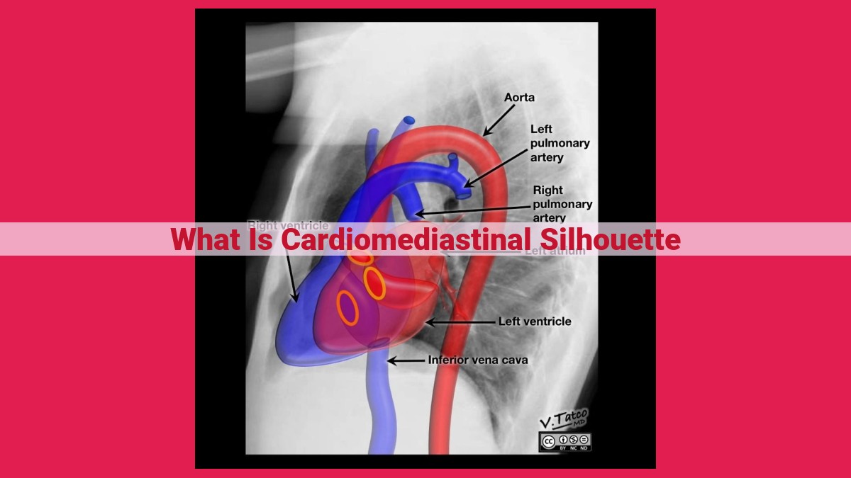
The cardiomediastinal silhouette is the heart-shaped outline visible on a chest X-ray. Its distinctive shape is formed by the silhouette of heart, major blood vessels, lymph nodes, and the thymus gland. The normal silhouette is well-defined and symmetrical, with clear borders and no unexpected enlargements or distortions.
Variations in the shape, size, or clarity of the cardiomediastinal silhouette can indicate abnormalities within the heart, lungs, or mediastinum. These abnormalities can stem from a variety of causes, including structural heart defects, lung diseases, mediastinal tumors, or infections. Understanding the types of abnormal silhouettes and their potential causes enables clinicians to narrow down the diagnostic possibilities and guide further investigations.
Types of Cardiomediastinal Silhouettes
Normal Cardiomediastinal Silhouette
The normal cardiomediastinal silhouette is a well-defined structure on a chest X-ray. It’s composed of the heart, aorta, and pulmonary vessels. The heart appears as a round or oval shape, occupying the left side of the silhouette. The aorta is a large blood vessel that carries oxygenated blood away from the heart, visible above the heart as a curved structure. The pulmonary vessels are smaller vessels that carry blood to and from the lungs, appearing as delicate branches extending from the heart.
Abnormal Cardiomediastinal Silhouettes
Abnormal cardiomediastinal silhouettes indicate potential underlying conditions. These abnormalities can range from changes in shape and size to the presence of additional structures. Some common abnormal silhouettes include:
- Cardiomegaly: Enlargement of the heart, often indicating heart disease or other underlying conditions.
- Aortic dilation: Enlargement of the aorta, which can be a sign of aortic aneurysms or other aortic disorders.
- Pericardial effusion: Accumulation of fluid around the heart, causing an enlarged and rounded silhouette.
- Mediastinal mass: Presence of a mass or tumor in the mediastinum, which can be a sign of cancer or other mediastinal conditions.
- Pulmonary edema: Fluid-filled lungs, resulting in a hazy appearance on the chest X-ray and an enlarged cardiomediastinal silhouette.
Causes of Abnormal Cardiomediastinal Silhouettes
The cardiomediastinal silhouette is a crucial indicator of heart and lung health. An abnormal silhouette can reveal underlying issues within the heart, lungs, or mediastinum, the area between the lungs. Let’s explore the various conditions that can lead to an abnormal cardiomediastinal silhouette:
-
Cardiovascular Causes: Heart failure occurs when the heart struggles to pump blood efficiently, leading to fluid buildup in the lungs, which may appear as an enlarged silhouette. Cardiomyopathy is a condition where the heart muscle is thickened or weakened, also resulting in an enlarged silhouette. Pericardial effusion occurs when fluid accumulates around the heart, causing the silhouette to appear “water bottle” shaped.
-
Pulmonary Causes: Conditions that affect the lungs can also alter the cardiomediastinal silhouette. Pneumonia is an infection that fills the lung air sacs with fluid, causing the silhouette to appear dense and hazy. Pneumothorax refers to collapsed lung tissue, leading to a smaller-than-normal silhouette on the affected side. Emphysema is a chronic lung disease that destroys lung tissue, creating areas of low density on the silhouette.
-
Mediastinal Causes: The mediastinum houses various structures, and abnormalities within these can impact the silhouette. Thymic hyperplasia is an enlargement of the thymus gland, often seen in children and young adults, resulting in a widened mediastinal shadow. Mediastinal masses can range from benign cysts to malignant tumors and may appear as a prominent mass on the silhouette. Aortic dissection is a life-threatening condition where the aorta tears, leading to an abnormal bulge in the silhouette.
Accurate Diagnosis and Management
Identifying the underlying cause of an abnormal cardiomediastinal silhouette is crucial for appropriate treatment. Detailed medical history, physical examination, and imaging tests, such as chest X-rays, CT scans, and echocardiograms, are essential for making an accurate diagnosis. Treatment varies depending on the specific cause and may involve medications, lifestyle changes, or surgical procedures. Timely diagnosis and management can significantly improve outcomes and ensure optimal cardiac and pulmonary health.
Diagnosis and Management of Abnormal Cardiomediastinal Silhouettes
Identifying an abnormal cardiomediastinal silhouette is crucial for timely diagnosis and effective management. Chest X-rays are the primary tool for evaluating the silhouette, revealing deviations from the normal shape and contours. Other imaging modalities like echocardiography, CT scans, and MRI provide detailed visualization of the heart and surrounding structures, aiding in accurate diagnosis.
Accurate diagnosis is paramount as it guides appropriate treatment. The underlying cause determines the course of action. For instance, congenital heart defects may require surgical intervention, while mediastinal tumors necessitate targeted therapy.
Treatment options are tailored to the specific condition. Heart failure may involve medications, lifestyle modifications, or device placement to support cardiac function. Lung disease often necessitates oxygen therapy, bronchodilators, or antibiotics to manage infections. Mediastinal masses may require surgical resection or radiation therapy.
Timely diagnosis and comprehensive management of abnormal cardiomediastinal silhouettes are essential for preserving cardiovascular health and overall well-being. By recognizing deviations from the normal silhouette and employing appropriate diagnostic tools, healthcare professionals can effectively identify and treat the underlying conditions, restoring patient comfort and improving long-term outcomes.