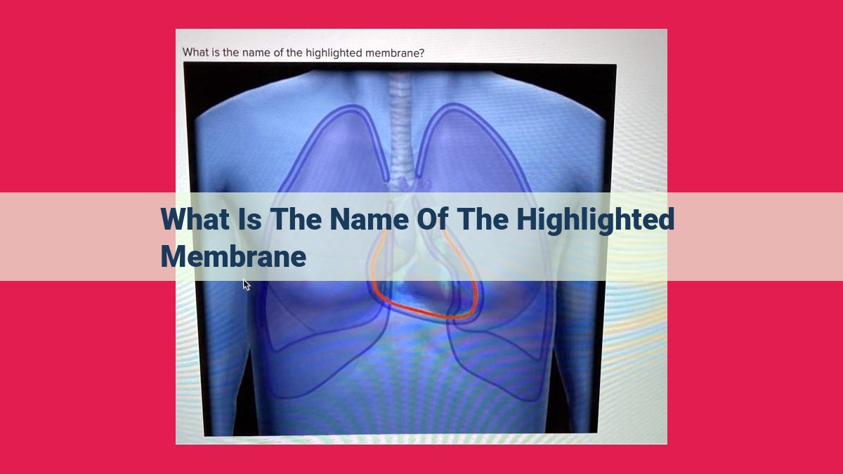
Basal Lamina: The Structural Foundation And Cell Signaling Hub Of Epithelial Tissues

The highlighted membrane is the basal lamina, a thin layer of extracellular matrix that lies beneath the plasma membrane of epithelial cells. It is composed of a dense network of collagen fibrils and proteoglycans, and it provides structural support and adhesion for the cells. The basal lamina is also involved in cell signaling and differentiation, and it plays a role in the development and maintenance of tissues.
Understanding the Basal Membrane: A Foundation of Cellular Biology
In the enigmatic world of cellular anatomy, the basal membrane holds a central stage. It’s a thin yet crucial layer that weaves a tapestry of support, dividing cells from the extracellular milieu.
The basal membrane’s significance lies in its gatekeeping role, regulating the flow of nutrients and signaling molecules essential for cellular function. It’s a testament to the intricate engineering of our bodies, enabling cells to thrive and communicate with precision.
Anatomy of the Basal Membrane: A Microscopic Gateway to Cellular Health
Beneath the bustling cityscape of cells, there lies a hidden realm—the basal membrane, a delicate yet crucial scaffold that governs the very way cells interact and function.
At the heart of the basal membrane beats the basement membrane, a thin but mighty layer composed of a labyrinthine network of proteins, carbohydrates, and lipids. Firmly anchored to the cells above and the extracellular matrix below, it acts as a bridge between two worlds, mediating communication and exchange.
Nestled within the basement membrane is the basal lamina, a 緻密 layer of specialized proteins that provides a firm foundation for cells to adhere. It’s like a sturdy rock upon which the cells can build their homes.
Flowing around the basal lamina and filling the gaps between cells is the extracellular matrix (ECM), a viscous yet pliable gel-like substance composed of various proteins and polysaccharides. This extracellular soup provides structural support, regulates cell growth and migration, and facilitates communication between cells and their surroundings.
As a testament to its importance, the basal membrane and its components play a symphonic role in maintaining tissue integrity and function. They provide anchorage for cells, ensuring they don’t float away like dandelion seeds in the breeze. They regulate the exchange of nutrients and waste products, keeping cells well-nourished and removing the clutter. And they orchestrate the signaling pathways that guide cells through their daily routines.
In the realm of cells, the basal membrane is more than just a supporting cast—it’s the backbone that holds everything together, enabling cells to thrive and tissues to flourish.
The Basement Membrane: A Closer Look
In the realm of cellular biology, the intricate architecture of the basement membrane plays a pivotal role in tissue function and cellular dynamics. It serves as an interface between cells and the surrounding extracellular matrix, orchestrating a symphony of cellular processes.
Relationship with Basement Membrane and Extracellular Matrix
The basement membrane is an integral part of the extracellular matrix, a complex network of proteins and carbohydrates that surrounds and supports cells. It lies beneath epithelial cells, forming a continuous sheet that delineates tissues and separates them from underlying connective tissue. The basement membrane consists of two distinct layers: the basal lamina and the reticular lamina.
Composition and Functions of the Basement Membrane
The basal lamina, also known as the basement membrane proper, is a thin layer directly adjacent to the plasma membrane of epithelial cells. It is primarily composed of type IV collagen, laminin, and proteoglycans. These components intertwine to form a latticework that provides structural support, allows for cell adhesion, and regulates cell proliferation and differentiation.
The reticular lamina lies above the basal lamina and is composed of a network of collagen fibers, primarily types III and IV. It connects the basement membrane to the underlying connective tissue and provides additional strength and flexibility.
Collectively, the basement membrane performs a myriad of functions, including:
- Cellular Adhesion: The basal lamina contains integrins, proteins that bind to receptors on the surface of epithelial cells, anchoring them to the basement membrane and providing structural stability.
- Tissue Separation: The basement membrane demarcated tissues, preventing the mixing of different cell types and maintaining tissue integrity.
- Filtration and Barrier: The extracellular matrix surrounding the basement membrane acts as a selective filter, allowing essential nutrients to reach cells while preventing potentially harmful substances from penetrating.
- Cellular Signaling: The basement membrane contains molecules that can interact with cell surface receptors, triggering intracellular signaling pathways that regulate cell behavior.
Basal Lamina: The Unsung Hero of Cellular Architecture
Beneath the bustling activity of cells lies a hidden world of intricate structures that form the foundation of our bodies. Among these structures, the basal lamina stands out as an unsung hero, playing a crucial role in maintaining tissue integrity and facilitating cellular communication.
A Closer Look at the Basal Lamina
The basal lamina is a thin, sheet-like layer that resides between epithelial cells and the underlying connective tissue. It’s composed of a meshwork of proteins, including collagen, laminin, and proteoglycans. These components interact to create a scaffold that provides structural support and regulates cell behavior.
The Glue That Binds Cells Together
One of the primary functions of the basal lamina is to anchor epithelial cells to the underlying connective tissue. It does this through specialized protein complexes called hemidesmosomes, which act as molecular glue, ensuring that cells remain firmly attached.
A Gatekeeper of Cellular Communication
Beyond its structural role, the basal lamina also serves as a gatekeeper of cellular communication. Its composition allows for the selective passage of nutrients and signaling molecules, controlling the flow of information between cells and the extracellular environment.
A Dynamic Landscape for Cellular Renewal
The basal lamina is a dynamic structure that undergoes constant remodeling, providing a foundation for cell growth and repair. During development, it guides cell migration and differentiation, shaping the specialized structures that define our tissues and organs.
As we journey through life, the basal lamina remains a silent guardian, ensuring that our cells function in harmony and maintain the delicate balance of our bodies. Its intricate architecture serves as a testament to the complexity and ingenuity of the human body, where even the smallest structures play a pivotal role in our well-being.
Extracellular Matrix: The Invisible Scaffold Shaping Cellular Life
Ensuring Structural Stability
Beneath the intricate dance of cells lies a hidden layer known as the extracellular matrix (ECM). This complex network of proteins and carbohydrates provides a firm foundation that supports and sculpts the very fabric of our tissues. Like an invisible scaffold, the ECM anchors cells, prevents tissue deformation, and provides physical cues for cell growth and differentiation.
Regulating Cellular Symphony
But the ECM’s role goes beyond structural support. It acts as a biochemical orchestra, regulating cell behavior and coordinating the symphony of intercellular communication. Glycosaminoglycans and proteoglycans, the main components of the ground substance, create an hydrated microenvironment that facilitates nutrient exchange and waste removal.
Guiding Cellular Decisions
The ECM also contains a variety of adhesive proteins, such as collagen and fibronectin. These proteins bind to cell surface receptors, transmitting signals that influence cell behavior. For instance, the ECM can promote cell adhesion, migration, and proliferation, playing a crucial role in tissue development and repair.
A Dynamic and Responsive Landscape
The extracellular matrix is not a static entity. It is dynamic and responsive, constantly remodeled to meet the changing needs of the tissue. Enzymes such as matrix metalloproteinases (MMPs) can break down the ECM, allowing for cellular reorganization and tissue remodeling.
A Keystone of Tissue Health
The extracellular matrix is an essential component of our tissues, providing structural support, regulating cellular behavior, and facilitating intercellular communication. Its dynamic nature allows tissues to adapt and respond to changes in their environment. Understanding the ECM is therefore crucial for unraveling the complexities of tissue biology and developing interventions for a wide range of diseases.
Ground Substance: The Matrix of Life
Nestled within the intricate tapestry of the extracellular matrix, the ground substance emerges as a vital player in the cellular symphony. Composed primarily of water, proteoglycans, and glycosaminoglycans, this semi-fluid matrix serves as a conduit for essential nutrients, facilitating cell growth and tissue repair.
Proteoglycans, adorned with long sugar chains, entrap water molecules to create a hydrated environment that nurtures cells. Glycosaminoglycans, the backbone of the ground substance, further contribute to its hydrophilic nature and provide mechanical support.
The ground substance is not merely a passive scaffold. It orchestrates a symphony of molecular interactions, regulating cell behavior. It harbors growth factors that stimulate cell division and migration, while simultaneously acting as a barrier to prevent uncontrolled cell growth.
Its viscoelastic properties cushion cells from mechanical stress, while its ability to bind water prevents tissue dehydration. The ground substance also plays a crucial role in immune responses, forming a protective barrier against pathogens.
In essence, the ground substance is the lifeblood of the extracellular matrix. It provides a supportive and nurturing environment that enables cells to thrive, orchestrates cellular communication, and safeguards tissues against damage.
Hemidesmosomes: Anchoring Cells to the Basal Membrane
Within the intricate cellular dance of life, the basal membrane plays a crucial role in organizing and supporting the cells that form our tissues. This thin yet remarkably strong layer is a cellular glue that holds epithelial cells firmly in place while allowing them to interact with their surroundings.
Hemidesmosomes are specialized structures that serve as the anchors between cells and the basal membrane. These protein complexes are composed of two primary components: transmembrane proteins that penetrate the cell membrane and bind to the basal membrane, and cytoplasmic proteins that connect to the cell’s cytoskeleton.
The transmembrane proteins of hemidesmosomes, such as integrins, act as molecular bridges, connecting the cell to the extracellular matrix (ECM), a web of proteins and other molecules that surrounds and supports cells. The ECM provides structural support and regulates cell behavior, ensuring that cells function in a coordinated and organized manner.
The cytoplasmic proteins of hemidesmosomes, such as plectin and desmoplakin, anchor the cell to the cytoskeleton, a network of filaments that provides structural stability and allows cells to move. This dual-anchorage system ensures that cells are firmly attached to both the basal membrane and the cytoskeleton, allowing them to withstand mechanical stresses and maintain their shape and position.
Hemidesmosomes play a vital role in cell adhesion and migration. They prevent cells from detaching from the basal membrane, maintaining the integrity of epithelial tissues. They also facilitate cell migration during development, wound healing, and certain pathological processes. Defects in hemidesmosomes can result in a variety of skin disorders and blistering diseases.
Understanding the structure and function of hemidesmosomes is essential for comprehending the mechanisms that regulate cell adhesion, migration, and tissue formation. These specialized structures play a critical role in maintaining the integrity of our tissues and enabling the proper functioning of our organs.