Differences In Atrial And Ventricular Walls For Optimal Heart Function

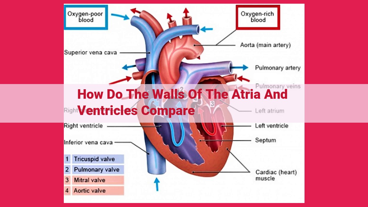
The walls of the atria are thinner and less muscular than those of the ventricles. This is because the atria are responsible for receiving and filling the ventricles with blood, while the ventricles must pump the blood out of the heart. The ventricular walls are thicker and more muscular to generate the necessary force for pumping. They contain trabeculae carneae, ridges of muscle that increase the surface area for contraction and enhance the force of the ventricular contractions.
Thickness: A Comparative Overview of Atrial and Ventricular Walls
The human heart is a remarkable organ responsible for pumping oxygenated blood throughout our bodies. It consists of four chambers: two atria and two ventricles. These chambers differ significantly in their thickness, reflecting their distinct functions.
The atrial walls are relatively thin, typically around 2-3 millimeters thick. This is because atria are mainly responsible for receiving blood from the body and the lungs. Their primary function is to fill the ventricles with blood, so they do not require the same level of muscular force as ventricles.
In contrast, the ventricular walls are much thicker, ranging from 5-10 millimeters in the left ventricle and 4-6 millimeters in the right ventricle. This increased thickness is necessary because ventricles are responsible for pumping blood out of the heart and into the bloodstream. They require a substantial amount of force to propel blood through the body’s arteries.
Muscularity: A Strength Comparison
The heart, an essential organ for life, is a complex and fascinating muscle. It pumps blood throughout the body, providing oxygen and nutrients to tissues and organs.
To accomplish this demanding task, the heart’s walls are composed of specialized muscle tissue. These muscles contract and relax in a coordinated rhythm, propelling blood through the circulatory system.
The ventricular walls, which form the lower chambers of the heart, are significantly more muscular than the atrial walls. This increased muscularity is crucial for generating the force necessary to pump blood out of the heart and into the body.
The muscles of the ventricular walls are arranged in a complex network of fibers that interweave and interlock. This intricate structure provides the strength and flexibility required for the powerful contractions that drive the heart’s pumping action.
In contrast, the atrial walls, which form the upper chambers of the heart, are thinner and less muscular. Their primary function is to receive blood from the body and fill the ventricles. As a result, they do not require the same level of force as the ventricles.
The difference in muscularity between the atrial and ventricular walls is a testament to the heart’s remarkable ability to adapt to its diverse functions. The thicker, more muscular ventricular walls provide the power to propel blood throughout the body, while the thinner, less muscular atrial walls facilitate the filling of the ventricles, ensuring a continuous flow of blood.
Trabeculae Carneae: The Heart’s Secret Weapon for Enhanced Contraction
Nestled within the muscular walls of the heart’s ventricles lie intricate structures called trabeculae carneae. These interconnected ridges and columns play a crucial role in maximizing the power of the heart’s contractions.
Imagine the ventricles as the powerhouses of the heart, responsible for pumping oxygen-rich blood throughout the body. To generate the necessary force for such a demanding task, the ventricular walls evolved to be thicker and more muscular than the atrial walls, which primarily receive and fill the heart with blood.
Within these muscular walls, trabeculae carneae serve as architectural marvels that dramatically increase the surface area of the ventricles. This increased surface area allows for a greater number of muscle fibers to be packed into the same space, further enhancing the heart’s contractile force.
But the benefits of trabeculae carneae extend beyond surface area. Their unique shape also plays a role. As the heart contracts, the trabeculae carneae interlock with each other, creating a more efficient and coordinated contraction.
Think of trabeculae carneae as the force multipliers of the heart. By increasing the surface area and improving the interlocking of muscle fibers, these structures enable the ventricles to generate the maximum force necessary to pump blood effectively throughout the body. Without trabeculae carneae, the heart’s pumping capacity would be significantly reduced, compromising the vital flow of oxygen and nutrients to our cells.
In summary, trabeculae carneae are remarkable adaptations that amplify the power of the heart’s contractions. They provide an ingenious solution to the challenge of generating sufficient force within the confined space of the ventricular walls. As a result, trabeculae carneae play an indispensable role in maintaining our cardiovascular health and ensuring the proper functioning of the entire body.
Ventricles: The Powerhouse of Pumping
As the heart’s lower chambers, the ventricles play a crucial role in pumping blood throughout the body. They are designed to generate the necessary force to propel blood through the arteries and into the body’s tissues.
The ventricular walls are considerably thicker and more muscular than those of the atria, the upper chambers of the heart. This enhanced muscularity allows the ventricles to generate the force required to pump blood efficiently.
Unique to the ventricles are structures called trabeculae carneae. These muscular ridges increase the surface area of the ventricular walls, allowing for more contractile force. The trabeculae carneae interdigitate with each other, creating a network that strengthens the ventricular walls and maximizes their contractile power.
The force generated by ventricular contractions is essential for maintaining blood circulation. The powerful contractions of the ventricles propel blood through the body’s arteries, delivering oxygen and nutrients to various organs and tissues.
In summary, the ventricles are the powerhouse of the heart, responsible for pumping blood throughout the body. Their thick, muscular walls and trabeculae carneae enable them to generate the necessary force for efficient blood circulation.
Atria: The Receiving and Filling Chambers of the Heart
When it comes to the life-sustaining miracle of the human heart, every chamber plays a crucial role. Amidst the rhythmic symphony of heartbeats, the atria, like celestial guardians, stand as the receiving and filling chambers, preparing the blood for its onward journey.
The atria, located at the heart’s upper reaches, serve as the gateways for blood returning from the body. Their walls, lined with smooth muscle, are thinner than those of the ventricles, allowing them to expand and accommodate the incoming blood. As the blood flows in, the atria swell, resembling two balloons awaiting their release.
Unlike their muscular counterparts below, the atria’s primary function lies in receiving and collecting. They act as reservoirs, holding the precious fluid until the ventricles are ready to pump it out. This filling phase, known as diastole, is essential for the heart’s coordinated operation, ensuring a steady supply of blood to the ventricles for their powerful ejections.
In this delicate dance of cardiac rhythm, the atria serve as the gentle conductors, preparing the blood for its vital mission. Their serene and spacious chambers provide a sanctuary for the weary blood as it gathers strength to fulfill its circulatory purpose. Without these receiving and filling chambers, the heart’s symphony would falter, and the life-giving flow of blood would cease.
Force: The Critical Element for Pumping
In the intricate tapestry of the human cardiovascular system, the heart holds a central stage, a tireless engine pumping life’s elixir through a labyrinth of blood vessels. Its two lower chambers, the ventricles, bear the brunt of this arduous task, generating the force that propels blood throughout our bodies.
The ventricular walls, composed of thick layers of muscle tissue, embody the heart’s power. This robust musculature is the foundation for vigorous contractions, the driving force behind blood circulation. The ventricles, like twin pistons, propel blood into the pulmonary artery and aorta, initiating its journey through the vascular network.
The strength of these contractions depends on the volume of blood filled into the ventricles. This volume, known as the preload, determines the degree of muscle fiber stretch and the subsequent force of contraction. A greater preload leads to more forceful contractions, while a reduced preload results in weaker ones.
The force of ventricular contractions is a crucial factor in maintaining blood pressure and ensuring adequate organ perfusion. Weak contractions can lead to hypotension, depriving tissues of vital oxygen and nutrients, while excessive force can strain the heart muscle and compromise its long-term health.
In summary, the force generated by ventricular contractions is the linchpin of blood circulation. This force, dependent on ventricular muscle thickness and preload, is paramount for maintaining blood pressure and sustaining the body’s vital functions.
Muscles: The Building Blocks of Cardiac Contraction
The human heart, a marvel of nature, is a tirelessly beating organ responsible for pumping oxygen-rich blood throughout our bodies. At the core of its exceptional function lies an intricate network of muscle tissue, the very essence of cardiac contraction.
Within the heart’s chambers, the walls are composed predominantly of muscle cells, interwoven in a highly organized manner. These specialized cells possess the remarkable ability to contract and relax in a synchronized rhythm, driving the heart’s pumping action.
The thickness and muscularity of the heart’s walls vary significantly, reflecting the distinct roles of the different chambers. The ventricles, the lower chambers responsible for pumping blood out of the heart, boast thicker and more muscular walls compared to the atria, the upper chambers that receive blood from the body. This disparity in muscle mass ensures that the ventricles can generate the force necessary to propel blood through the body’s extensive network of blood vessels.
The inner surfaces of the ventricles are adorned with trabeculae carneae, muscular ridges that further enhance the heart’s pumping efficiency. These interconnected columns increase the surface area of the ventricles, providing more attachment points for muscle fibers. As the ventricles contract, the trabeculae carneae pull inward, maximizing the force generated and optimizing the expulsion of blood.
In conclusion, the heart’s muscle tissue forms the foundation for its remarkable ability to pump blood. The interplay of thickness, muscularity, and trabeculae carneae ensures that the heart can generate the force necessary to sustain life, delivering essential oxygen and nutrients to every corner of our bodies.
Contraction: The Rhythmic Heartbeat
The human heart, a remarkable organ, is responsible for pumping oxygenated blood throughout the body, supplying every cell with the life-sustaining nutrients. At the core of this essential function lies the process of ventricular contraction, a rhythmic symphony of muscular movement that powers the heart’s pumping action.
The ventricles, the heart’s lower chambers, are thicker and more muscular than the atria, the upper receiving chambers. This architectural difference reflects their distinct roles. The ventricles’ task is to propel blood out of the heart with sufficient force to reach all corners of the body’s vast vascular network.
To achieve this forceful ejection, the ventricular walls are composed of intricately arranged muscle cells. These muscle fibers are organized into spiraling bands that crisscross and interlock, creating a robust and dynamic contractile apparatus.
Further enhancing the ventricular walls’ contractile power are specialized muscular ridges known as trabeculae carneae. These ridges project inward from the ventricular walls, increasing their surface area and providing attachment points for the muscle fibers. The increased surface area allows for a greater number of muscle fibers to contract simultaneously, maximizing the force generated.
As an electrical impulse initiates a heartbeat, it travels through the heart’s conduction system, triggering the contraction of the ventricles. The muscle fibers of the trabeculae carneae, along with those of the ventricular walls, contract vigorously, exerting an inward force that decreases the ventricular volume. As the ventricular volume diminishes, the blood within them is forcefully expelled out of the heart and into the body’s arteries, initiating the next vital cycle of oxygen and nutrient delivery.
Thus, the ventricular contraction, meticulously orchestrated by the synchronized contraction of its muscular walls and trabeculae carneae, serves as the heart’s pumping engine, driving the rhythmic flow of life-giving blood throughout the body.